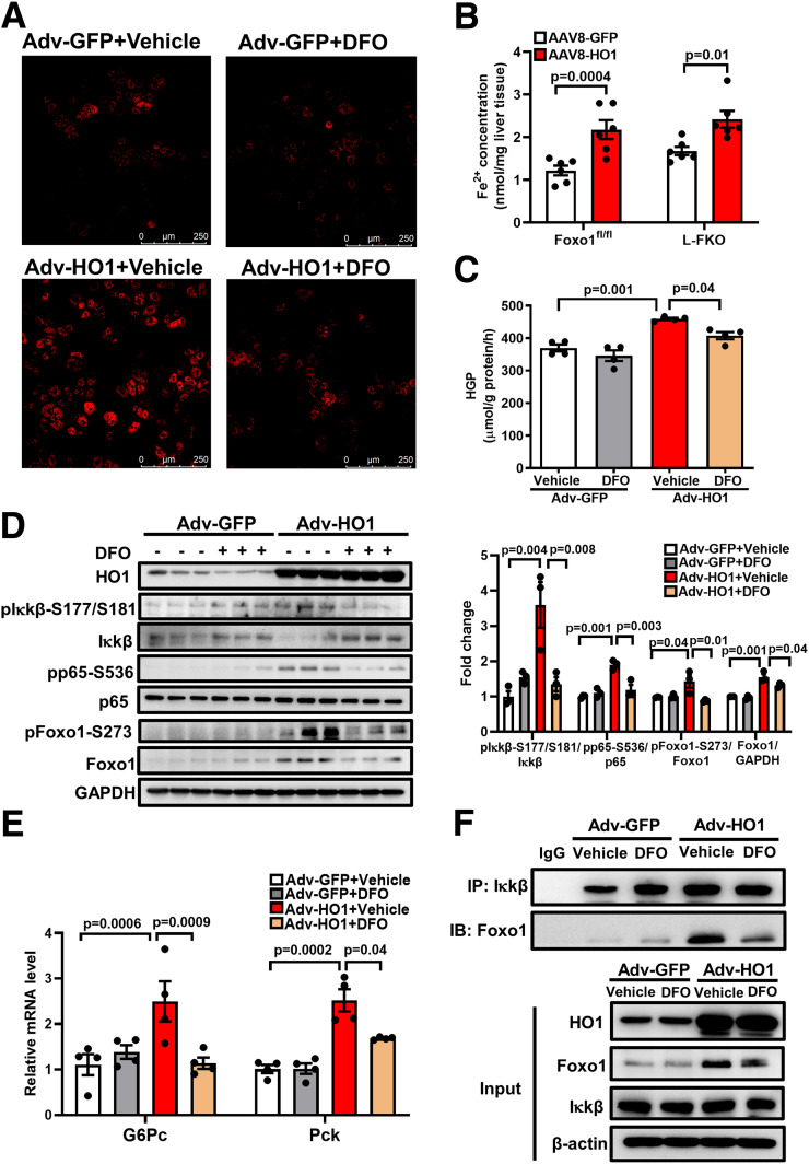
Figure 5.
HO1 overexpression activates hepatic NF-κB and Foxo1 by generating ferrous iron. A: Hepatocytes from wild-type mice were isolated and transfected with Adv-GFP or Adv-HO1 for 22 h and then treated with 40 μmol/L DFO for 2 h. The FeRhoNox-1 probe was used to detect the intracellular ferrous iron, which was visualized by confocal microscope. B: AAV8-GFP or AAV8-HO1 was delivered via retro-orbital to male mice (8–12 weeks old). Tissues from the mice were collected 14 days after the AAV8 injection. The concentration of ferrous iron in the liver tissue was measured and normalized by the weight of liver used for the assessment. n = 6 mice/group. Results are presented as mean ± SEM. C: Hepatocytes from wild-type mice were isolated. The cells were pretreated with 40 μmol/L DFO for 0.5 h and then transfected with Adv-GFP or Adv-HO1 for 20 h. Afterward, the cells were switched to HGP buffer for 3 h. HGP was normalized to total protein levels. n = 4/group. Results are presented as mean ± SEM. D: Hepatocytes from wild-type mice were isolated. The cells were pretreated with 40 μmol/L DFO for 0.5 h and then transfected with Adv-GFP or Adv-HO1 for 20 h. The cell lysates were loaded onto a 10% SDS-PAGE for Western blotting analyses to detect HO1, pIκkβ-S177/S181, total Iκkβ, pp65-S536, total p65, pFoxo1-S273, and total Foxo1. The levels of phosphorylated proteins were normalized to the corresponding total protein level. The level of total Foxo1 was normalized to GAPDH. n = 3/group. Results are presented as mean ± SEM. E: Hepatocytes from wild-type mice were isolated. The cells were pretreated with 40 μmol/L DFO for 0.5 h and then transfected with Adv-GFP or Adv-HO1 for 24 h. The RNA was extracted and expression of G6Pc and Pck determined by quantitative PCR. The cellular protein lysates from E were used for immunoprecipitation of Iκkβ. F: The immunoprecipitation (IP) complex and the cell lysates (input) were loaded onto a 10% SDS-PAGE for Western blotting analyses. IB, immunoblotting.

