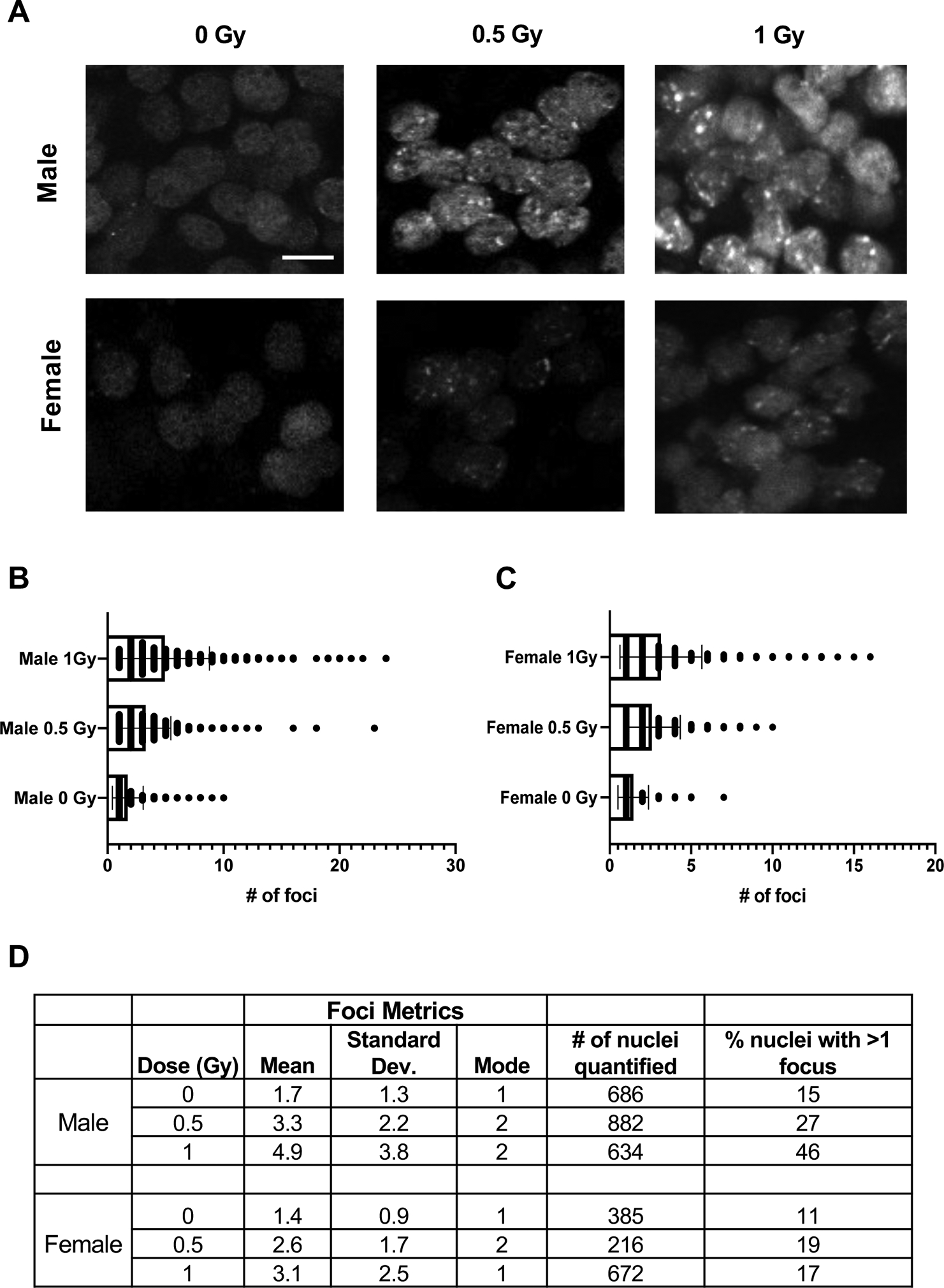
Figure 2. Primordial germ cells of Oct4ΔPE 53BP1-mCherry transgenic mice form radiation-induced foci in a dose-dependent manner.

(A) Whole mount immunofluorescence images of E13.5 transgenic male and female genital ridges 1 hour after exposure to 0, 0.5, or 1 Gy of radiation. Scale bar=10 μM. (B) Foci quantification of transgenic male PGCs at the doses shown in A; p-values derived from unpaired Student’s t-tests where p<0.0001 for all comparisons shown. (C) Foci quantification of transgenic female PGCs at the doses shown in A; p-values derived from unpaired Student’s t-tests where p=0.002 for the 0.5 vs. 1 Gy statistical comparison and p<0.0001 for the 0 vs. 0.5 Gy and 0 vs. 1 Gy comparisons. (D) Table of reporter induction metrics in response to radiation including mean number of foci formed, mode, number of nuclei quantified per condition and the percentage of nuclei with more than one focus.
