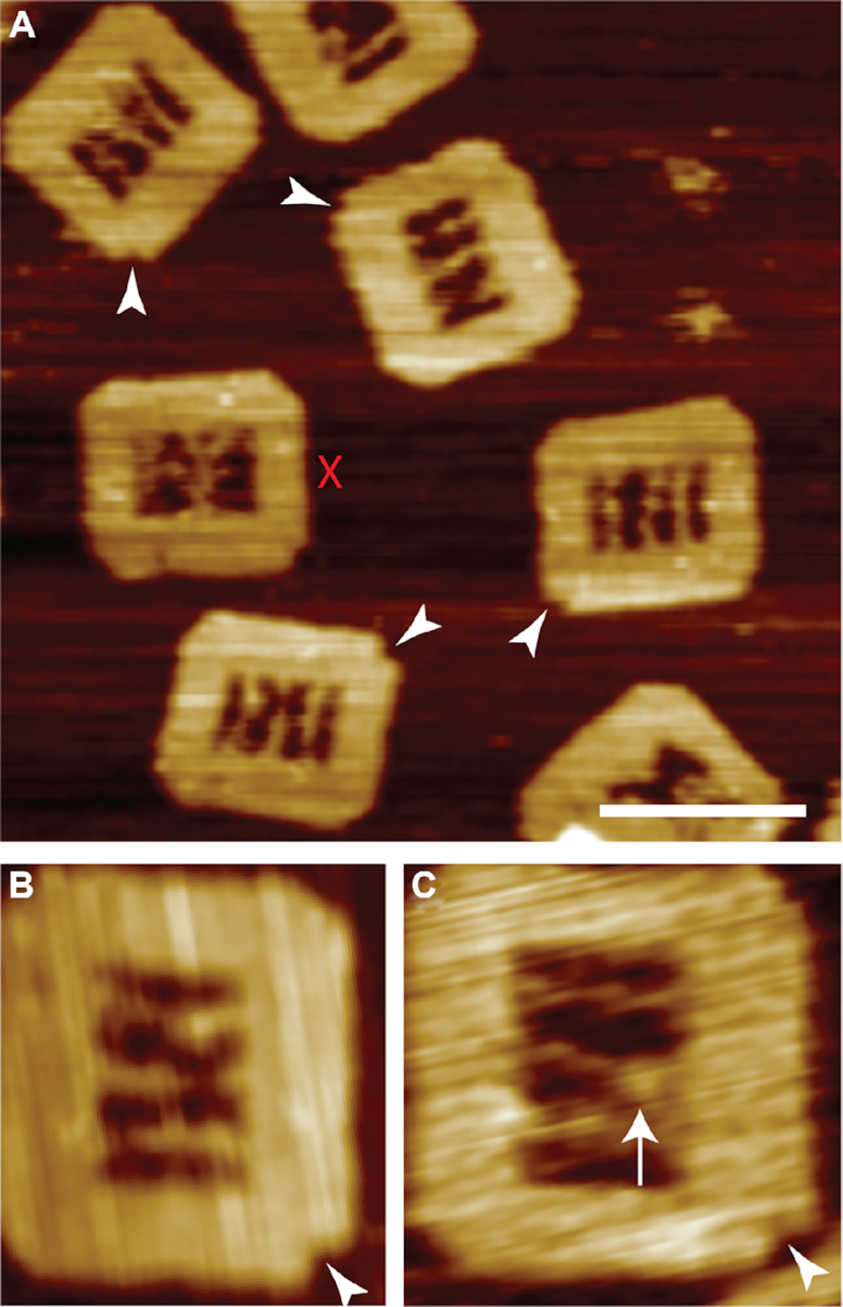
Figure 2.

Assembly of the DNA frame and formation of RecA nucleoprotein filament on the NPF DNA. (A) AFM images of correctly assembled DNA frames including successful incorporation of all three internal DNA molecules. The polarity markers are indicated (white triangles). DNA frames with ambiguous arrangements of the internal DNA molecules are discounted (red cross) from subsequent analyses. Individual DNA origami frames are shown prior to (B) and following RecA nucleoprotein filament formation (C). The nucleoprotein filament is clearly visible on the ssDNA region (white arrow). Scale bar = 100 nm.
