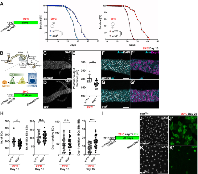
Figure 1.
Loss of ecd shortens lifespan and compromises maintenance of the adult intestine. (A) Control w1118 and temperature-sensitive ecd1 fly lines were allowed to develop at 18°C. After eclosion and two-day mating, males and females were separated and upshifted to restrictive temperature of 29°C. Lifespan curves show percentage of survival of w1118 and homozygous ecd1 adult males and females over time (n = 300) at 29°C and represent one of the two independent experiments. Adult ecd1 male and female flies were shorter lived (mean difference of 6.1 days for males and 10.2 days for females) compared to control flies. Statistical significance was determined by a log-rank test, ***P < 0.001. (B) Schematic diagram of Drosophila adult intestine. Posterior midgut is maintained by the intestinal stem cells (ISCs), which divide asymmetrically to self-renew and generate either the postmitotic enteroblasts (EBs) or the enteroendocrine (EE) lineage precursors. The midgut ISCs and EBs express the transcription factor Escargot (Esg) while EEs are positive for Prospero (Pros). EBs further differentiate into large absorptive enterocytes (ECs) characterized by polyploid nuclei and expression of Myosin 1A (Myo1A). Schematics of experimental setup and timeline to assess impact of ecd deficiency on adult midgut. (C, D) Representative confocal micrographs show that posterior midguts of ecd1 homozygous mutant flies (D) kept at restrictive temperature for 15 days are thinner compared to controls (w1118) (C). Scale bars: 50 μm. (E) Diameter measurements of posterior midguts from control (w1118, n = 8) and ecd1 flies (n = 12) kept at 29°C for 15 days. Nuclei were stained with DAPI. Statistical significance was determined by unpaired two-tailed Student's t-test. Data represent means ± SD, **P < 0.01. (F, G) Representative confocal micrographs of posterior midguts from adult female flies kept at restrictive temperature for fifteen days show increased number of apoptotic Dcp-1-positive cells (magenta) in ecd1 (G, G’) compared to control (w1118) intestines (F, F’). Cell membranes were visualized by Armadillo (Arm) antibody (cyan). Note the stronger Arm signal highlighting smaller progenitors compared to weaker staining of large polyploid ECs (F’, G’). DAPI stains nuclei. Scale bars: 50 μm. (H) Cell counts and quantification of Dcp-1-positive cells revealed reduced number of ECs and elevated apoptosis in progenitor and EE population in posterior midguts of ecd1 adult female flies (n = 34) kept at 29°C for 15 days compared to control (w1118, n = 30). Unpaired nonparametric two-tailed Mann–Whitney test was used to calculate P-values. Data represent means ± SD, **P < 0.01, ****P < 0.0001, n.s. = non-significant. (I) Experimental setup and timeline for RNAi-mediated silencing of ecd in progenitors (ISCs and EBs) of adult midguts using esgTS> expression system. (J, K) Representative confocal images of twenty-day-old control posterior midguts (J) and those expressing ecdRNAi (K) in ISCs/EBs (esgTS>) marked by GFP show decrease in Esg-positive progenitor cells following ecd knockdown (compare J’ and K’). DAPI stains nuclei. Scale bars: 50 μm. See also Supplementary Figure S1.

