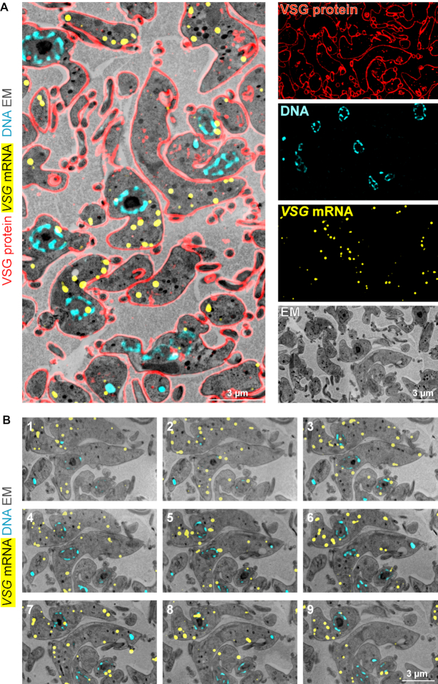
Figure 6.
Combination of LR White smFISH with immunofluorescence and array tomography. (A) LR White embedded BSF trypanosomes were probed for VSG mRNA (shown in yellow, imaged by standard fluorescence microscopy), followed by antibody incubation with anti-VSG (red, imaged by structured illumination microscopy) and DNA staining with DAPI (cyan). After imaging, samples were contrasted and imaged on a scanning electron microscope. Correlated images and individual images are shown. (B) Samples from (A) were cut into 41 sections, with 100 nm thickness. All slices were probed for VSG mRNA, stained with DAPI for DNA and imaged by fluorescence microscopy. Samples were contrasted, imaged by scanning electron microscopy and assembled to a stack. Nine images of the stack are shown; for the entire stack see Supplementary movie S1.

