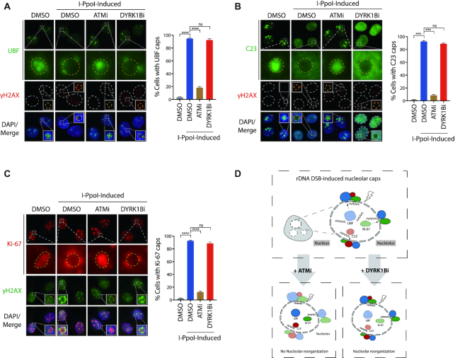
Figure 2.
Uncoupling of ATM-dependent nucleolar cap formation. (A–C) U2OS I-PpoI cells were pre-treated with ATM inhibitor (ATMi; KU-55933) or DYRK1B inhibitor (DYRK1Bi; AZ191) for 1 h before supplementation with Shield-1 and 4-OHT for 4 hr. After fixation, cells were processed for immunofluorescence with anti-UBF (A), anti-C23 (B), anti-Ki-67 (C) or anti-γH2AX antibodies, respectively. The squares show the enlarged nucleoli and the dashed circles indicate margins of the enlarged nucleoli. Percentages of cells with the indicated nucleolar caps were quantified from at least two experiments and 300 cells from each condition were analysed. Bars represent mean ± SEM; ns, not significant; ***P< 0.001; ****P< 0.0001. (D) Graph illustrates rDNA DSBs-induced nucleolar reorganization following ATM or DYRK1B inhibition.

