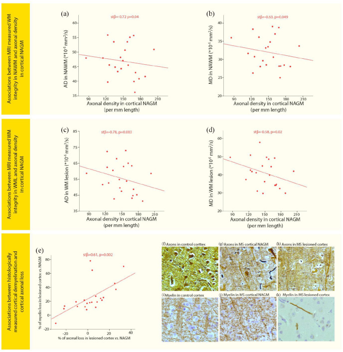
Figure 3.
Associations of WM pathology and cortical pathology with cortical axonal loss. Scatter plots displaying associations between (a) axial diffusivity (AD) in normal-appearing white matter (NAWM) and axonal density in cortical normal-appearing gray matter (NAGM) in MS, (b) Mean diffusivity (MD) in NAWM and axonal density in cortical NAGM in MS, (c) AD in white matter lesions (WML) and axonal density in cortical NAGM in MS, (d) MD in WML and axonal density in cortical NAGM in MS, (e) the percentage reduction in axonal density in lesioned cortex compared with cortical NAGM and percentage reduction in myelin density in lesioned cortex compared with cortical NAGM. Images show axonal staining (Bielschowsky) in (f) control cortex, (g) MS cortical NAGM, and (h) MS lesioned cortex. Images showing myelin staining (PLP) in an adjacent tissue section and same location as the axonal pictures were taken in (i) control cortex, (j) MS cortical NAGM, and (k) MS lesioned cortex.

