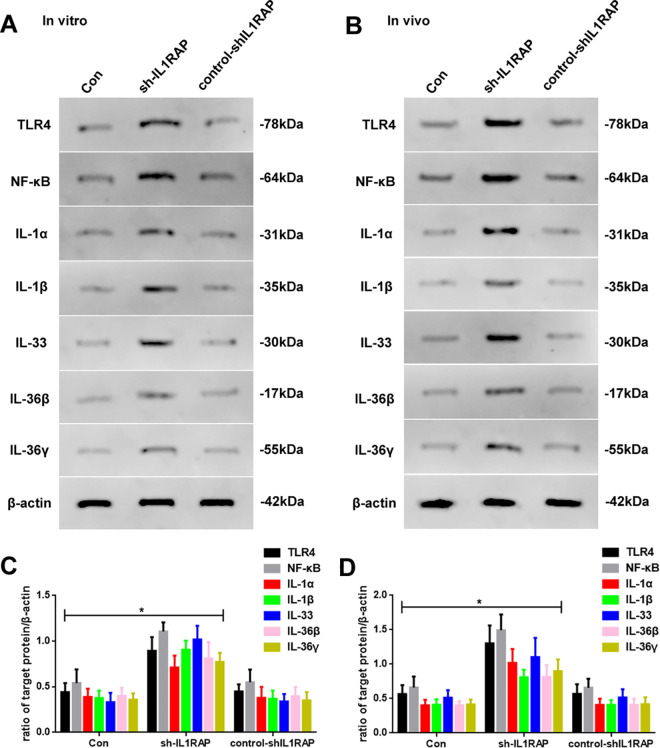
Figure 5.
(A) Western blot assay of TLR4, NF-κB, IL-1α, IL-1β, IL-33, IL-36β, and IL-36γ expression of Con, sh-IL1RAP, and control-shIL1RAP groups in vitro. (B) Western blot assay of TLR4, NF-κB, IL-1α, IL-1β, IL-33, IL-36β, and IL-36γ expression of Con, sh-IL1RAP, and control-shIL1RAP mice in vivo. (C) Quantification of TLR4, NF-κB, IL-1α, IL-1β, IL-33, IL-36β, and IL-36γ of Con, sh-IL1RAP, and control-shIL1RAP groups in vitro. (D) Quantification of TLR4, NF-κB, IL-1α, IL-1β, IL-33, IL-36β, and IL-36γ of Con, sh-IL1RAP, and control-shIL1RAP groups in vivo. Protein levels were normalized to β-actin (sh-IL1RAP vs. other group, * P < 0.05, n = 6 per group).

