Figure 1.
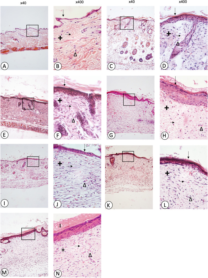
Microphotographs of skin sections of wounds in normal mice: skin without wound (A, B), control (C, D), vehicle (E, F), Recoveron (G, H), latex 50% (I, J), latex 75% (K, L), and latex 100% (M, N). Hematoxylin and eosin staining, Original increase and area framed to ×400; epidermis ( ↓), dermis (+), collagen fibres ( → ), fibroblasts (Δ).

