Fig. 1.
Osteogenesis imperfecta bone tissues assessed by light microscopy photomicrographs are shown.
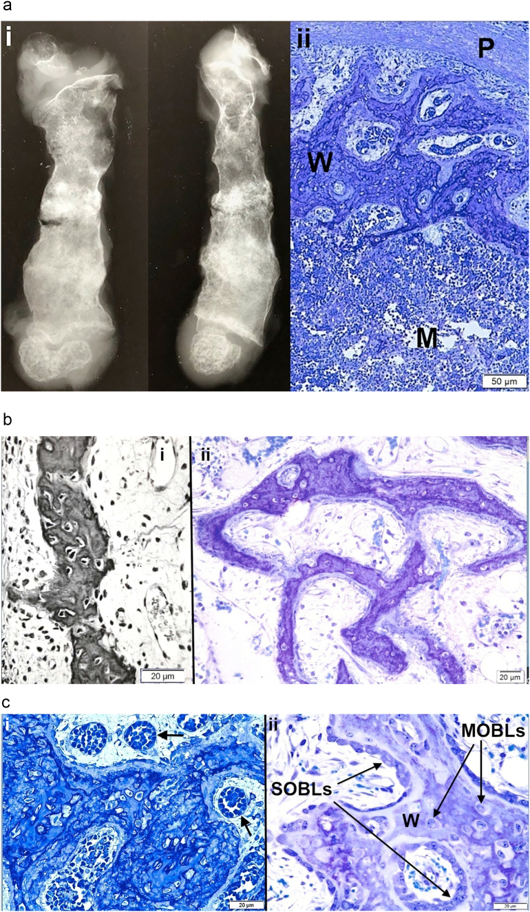
a. i) Anteroposterior and lateral views of a femoral specimen X-ray from an OI Sillence II (OIC A) patient who died a few weeks after birth are shown; ii) Cross-section of the femur diaphysis (same specimen) shows periosteum (P) at top, a narrow collection of discontinuous cortical woven bone (W) and an extensive marrow cavity (M) without trabeculae below. Plastic embedded 1% toluidine blue stained section.
b. i) Woven bone from tibial cortex of a type II (OIC A) patient is surrounded by cellular tissue with vessels. Paraffin embedded hematoxylin and eosin stained (black and white) section. ii) A separate section of tibial cortex in a type II (OIC A) patient shows thin woven bone spicules separated by wide minimally cellular spaces. Plastic embedded 1% toluidine blue stained section.
c. i) Woven bone alone is present in femoral cortex from type II (OIC A) patient. The tissue is well vascularized (arrows). Woven bone cells are round to oval and closely packed. Plastic embedded 1% toluidine blue stained section. ii) Woven bone (W) accumulations, even in type II bone, when sufficiently extensive can lead to coverage by surface osteoblasts (SOBLs). MOBLs = mesenchymal osteoblasts. Plastic embedded 1% toluidine blue stained section.
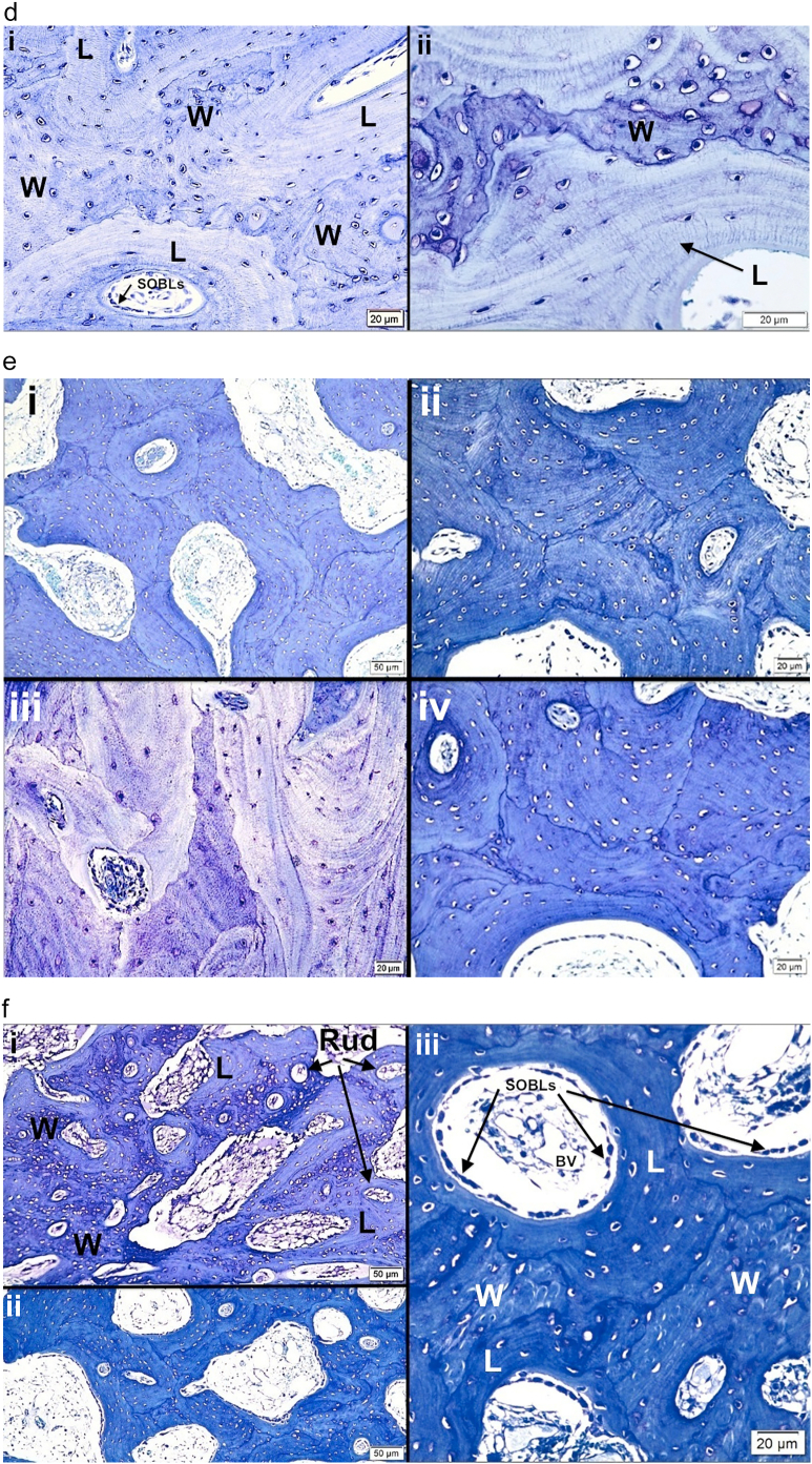
d. i) Lamellar on woven bone pattern of tissue deposition is seen with close observation in specimen from type III patient. Cell shape is distinctive in each tissue conformation; woven bone cells are round to oval and lamellar bone osteocytes are elliptical and elongated along the long axis of the lamellae. W = woven bone, L = lamellar bone and SOBLs = surface osteoblasts. Plastic embedded 1% toluidine blue stained section. ii) Higher power LM shows lamellar (L) on woven (W) bone deposition in specimen from type III patient. Plastic embedded 1% toluidine blue stained section.
e. Four photomicrographs (i-iv) from tibial cortical bone in different type III patients demonstrate the mosaic pattern of lamellar OI bone formation in severe to moderate cases. The lamellae are short and obliquely positioned to one another. Figures i, ii and iv are also examples of partial compaction of the forming lamellar bone. Plastic embedded 1% toluidine blue stained sections.
f. i) Rudimentary (Rud) osteon formation begins as lamellar (L) bone on woven (W) bone formation increases. Tissue shown is from a severe type III (OIC B) femur. Plastic embedded 1% toluidine blue stained section. ii) The amount of lamellar bone is greatly increased in this type III patient and compaction has increased from rudimentary to partial. Plastic embedded 1% toluidine blue stained section. iii) Section illustrates the mechanism of osteonal formation from SOBLs synthesizing lamellar tissue and closing in on longitudinal blood vessels (BV). Lamellar (L) tissue has been deposited on woven (W) bone scaffolds in type III patient. Plastic embedded 1% toluidine blue stained section.
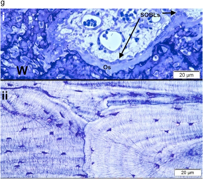
g. i) Osteoid (Os) tissue layer is prominent in this type II tissue section. The osteoid has been synthesized by the SOBLs on the woven (W) bone scaffold. Plastic embedded 1% toluidine blue stained section. ii) Lamellar bone from a type III patient shows a dense concentration of canaliculi linking adjacent osteocytes. Plastic embedded 1% toluidine blue stained section.


