Fig. 2.
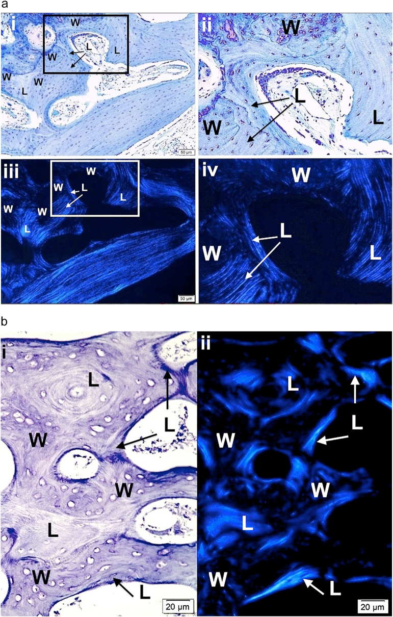
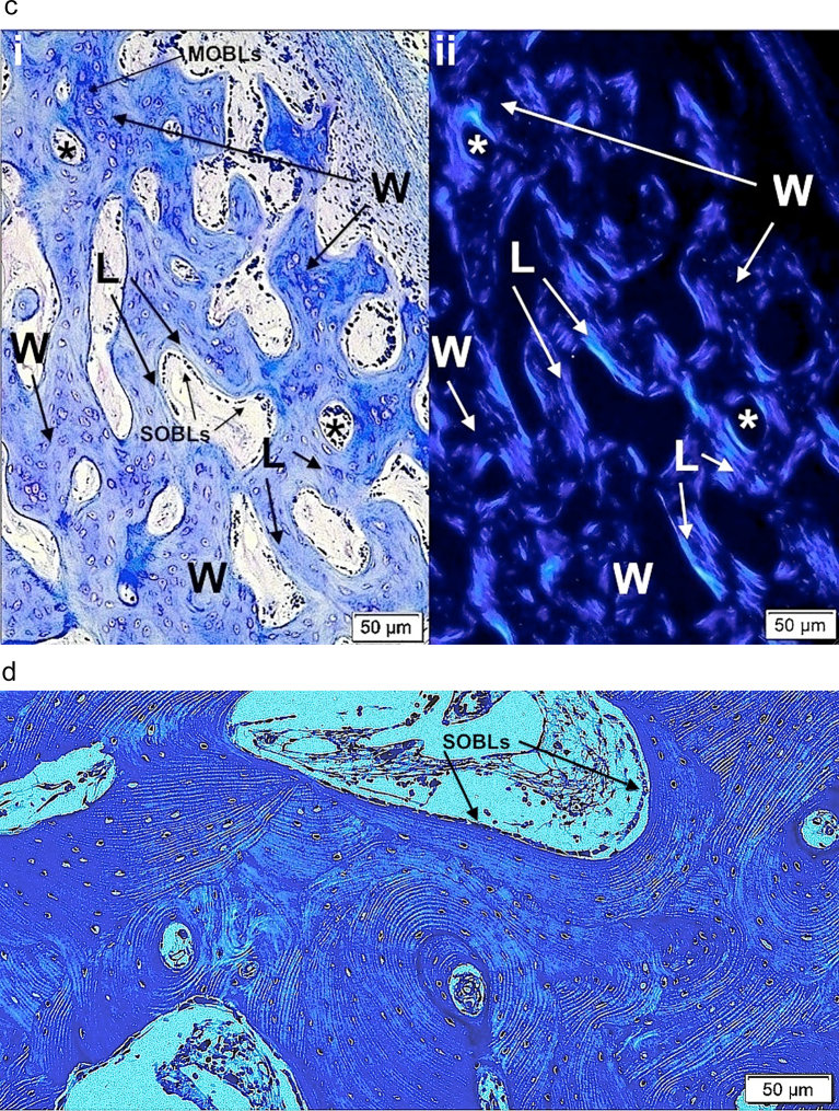
Images show the value of using polarizing light microscopy (PLM) to highlight woven and lamellar conformations in osteogenesis imperfecta bone.
a. i) and ii) LM photomicrographs shows both lamellar and woven bone from a type III patient. The box in i outlines the region magnified in ii. Woven (W) bone is relatively hypercellular with round to oval cells compared with lamellar (L) bone where cells are elliptical and oriented along the axis of the lamellae. iii) and iv) PLM views show the same tissue seen by LM in i and ii. The box in iii outlines the region magnified in iv. Woven (W) bone regions are dark due to the random orientation of the collagen fibrils producing an isotropic effect with polarization. Lamellar (L) bone regions are light due to the parallel orientation of the fibrils producing an anisotropic effect allowing passage of light with polarization. The clear-cut differentiation of woven and lamellar tissue is highlighted by the PLM technique. Plastic embedded 1% toluidine blue stained section.
b. i) LM section of cortical bone from patient with type II OI shows mixture of woven (W) and lamellar (L) bone. At this magnification one can distinguish the 2 conformations, especially by looking at the cellularity regarding number and shape. ii) PLM view of the exact same section highlights lamellar (L) bone as light blue (anisotropic) and woven (W) bone as dark (isotropic). Thin short streaks of light blue within woven areas represent the earliest phases of lamellar bone deposition on a sufficient woven scaffold. Plastic embedded 1% toluidine blue stained section.
c. i) A section of femoral bone assessed by LM is shown; ii) the same section is assessed by PLM. MOBLs = mesenchymal osteoblasts synthesizing woven (W) bone and SOBLs = surface osteoblasts synthesizing lamellar (L) bone. An * (asterisk) = central vascular canal of forming osteon. Plastic embedded 1% toluidine blue stained section.
d. Section of mosaic pattern in OI diaphyseal bone using partial polarization allows for cellular visualization as well as lamellar matrix orientation. The bone tissue is all lamellar and partially compacted but shows multiple short lamellae in variable orientations (cross-section, longitudinal and oblique). With rotation of the slide, regions appearing dark show the parallel lamellar orientation. Plastic embedded 1% toluidine blue stained section under partial polarization.


