Fig. 6.
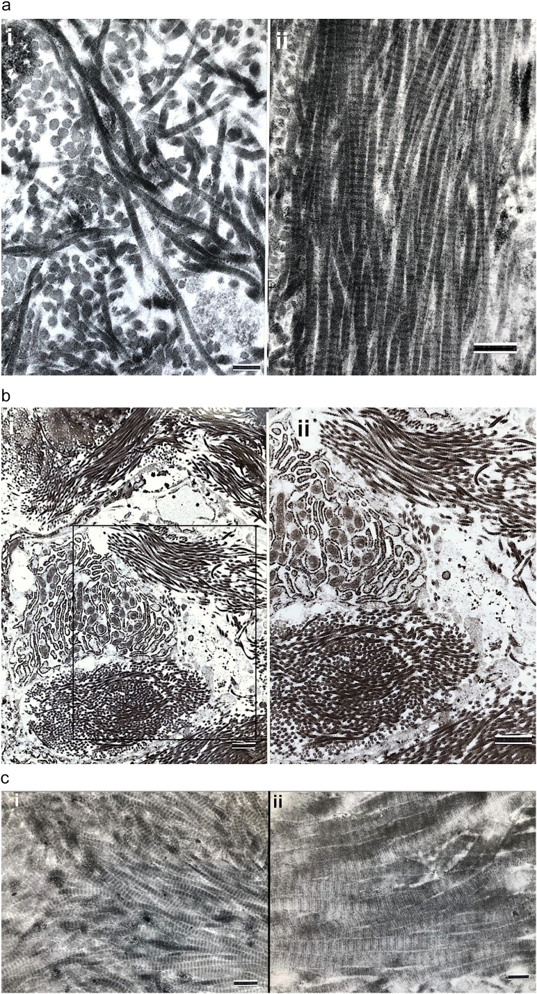
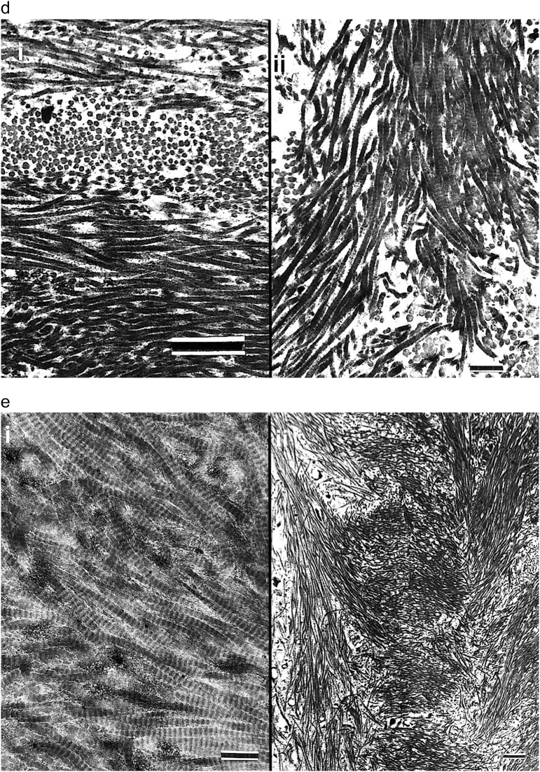
Transmission electron micrographs of the collagenous matrix in OI woven and lamellar bone are shown.
a. i) Fibrils from woven bone in a severe type III (OIC B) patient show characteristic random fibril orientation. Marker = 0.22 μm; ii) Fibrils from lamellar bone in a different patient with severe type III OI (OIC B) are unidirectional. Marker = 0.38 μm.
b. MOBL (center left) in woven bone tissue in a patient with type III OI shows markedly dilated RER. The cell is surrounded by 6 prominent bundles of unidirectional collagen fibrils but the bundles are obliquely positioned (randomly oriented) to one another. Marker = 1.08 μm. ii) Tissue from the box region in i) is magnified in ii) to highlight the fibrillar bundle random orientation. Marker = 1.08 μm.
c. Fibrils from lamellar bone in two patients both with severe type III OI are shown. i) Collagen fibrils show unidirectional orientation at right and a clear tendency to gradual deviation in direction upwards and downwards at left. Marker = 0.45 μm; ii) Collagen fibrils with unidirectional pattern also tend to a gradual angular deviation. The fibrils show the characteristic collagen crossbanding at this TEM magnification. Marker = 0.19 μm.
d. i) Orthogonal array of collagen fibril deposition in adjacent layers in lamellar bone is readily seen at these magnifications. Patient had a severe type III OI. Marker = 0.76 μm; ii) Collagen fibrils in lamellar bone in a severe type III (OIC B) patient show unidirection at top and angular deviation at bottom. Marker = 0.55 μm.
e. TEM images are shown from lamellar bone in two patients with type III OI. i) Unidirectional collagen fibrils are present but a gradual angular deviation as great as 45° is seen. Marker = 0.45 μm; ii) A well-organized fibrillar weave (herringbone pattern) characterizes some sections of lamellar bone. Marker = 0.93 μm.


