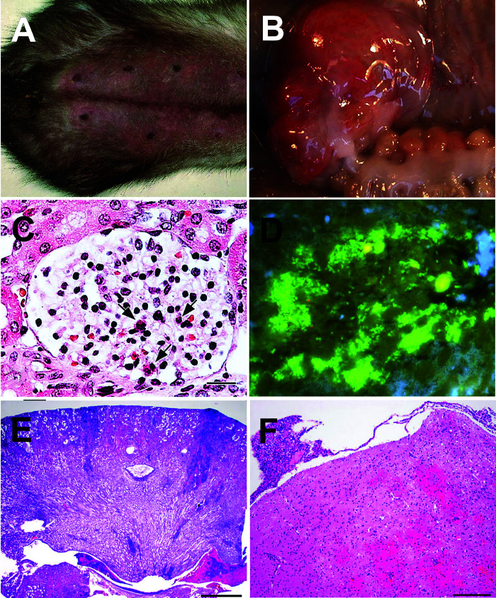
Figure 12.
(A) Ferret naturally infected with E. coli exhibiting signs of mastitis including swollen and erythematous mammary tissue. (B) Hyperemic and hemorrhagic serosa at the level of the distal cecum adjacent to the junction with the proximal colon in a Dutch Belted rabbit experimentally infected with enterohemorrhagic E. coli O157:H7; Copyright © American Society for Microbiology, [Infection and Immunity 80, pages 369-380, 2012]. (C) Global intracellular edematous swelling, increased numbers of heterophils (arrows), and decreased number of erythrocytes (“bloodless glomerulus”) in a glomerulus of a Dutch Belted rabbit experimentally infected with enterohemorrhagic E. coli O153 (scale bar: 60 µm); García and colleagues, Renal Injury Is a Consistent Finding in Dutch Belted Rabbits Experimentally Infected with Enterohemorrhagic Escherichia coli, The Journal of Infectious Diseases, 2006, volume 193, issue 8, pages 1125-1134, by permission of the Infectious Diseases Society of America. (D) E. coli-associated necrotizing suppurative metritis (pyometra) in a naturally infected “alpha V integrin+/-; alpha v fl/+; Tie 2, Cre+/-” mouse. E. coli is fluorescently labeled with a green peptic nucleic acid in situ hybridization probe that detected bacteria in the affected and luminal areas of the uterus. The nuclei of the cells are stained blue with 4’,6’-diamidino-2-phenylindole (DAPI) (no scale bar: x100); Reprinted from Microbes and Infection18(12), García A, Mannion A, Feng Y, Madden CM, Bakthavatchalu V, Shen Z, Ge Z, Fox JG, Cytotoxic Escherichia coli strains encoding colibactin colonize laboratory mice, 777-786, Copyright (2016) with permission from Elsevier. (E) Renal section of a mouse naturally infected with cytotoxic E. coli (pks+) and exhibiting multifocal subacute suppurative pyelonephritis, intraluminal bacteria, and tubular necrosis (scale bar: 1 mm). (F) Brain section of a mouse naturally infected with cytotoxic E. coli (pks+) and exhibiting focally extensive subacute necrohemorrhagic meningoencephalitis (scale bar: 200 µm). Figures 12(E) and 12(F) have been reprinted from Bakthavatchalu and colleagues (2018) Cytotoxic Escherichia coli strains encoding colibactin isolated from immunocompromised mice with urosepsis and meningitis. PLoS One 13(3): e0194443. doi: 10.1371/journal.pone.0194443, with permission through an open access Creative Commons Attribution (CC BY) license. (C, E, F are hematoxylin and eosin stained sections).

