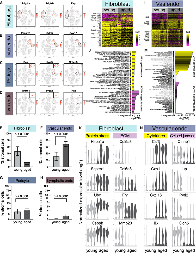
Figure 6. Age-Dependent Alterations in Stromal Fibroblasts and Vascular and Lymphatic Cells.
(A–D) Fibroblasts (A), vascular endothelial cells (B), pericytes (C), and lymphatic endothelial cells (D) are distinguished by characteristic makers in scRNA-seq data.
(E–H) Relative proportions of fibroblasts (E), vascular endothelial cells (F), pericytes (G), and lymphatic endothelial cells (H) in young (n = 3) and aged (n = 4) mice by scRNA-seq (Fisher’s exact test).
(I) Differentially expressed genes in young versus aged fibroblasts. See also Table S3.
(J) Top gene sets identified by pathway analysis of differentially expressed genes in (I), with recurring gene sets highlighted. See also Table S4.
(K) Violin plots of select genes in (I) and (J) for fibroblasts.
(L) Differentially expressed genes in young versus aged vascular endothelial cells. See also Table S3.
(M) Top gene sets identified by pathway analysis of differentially expressed genes in (L), with recurring gene sets highlighted. See also Table S4.
(N) Violin plots of select genes in (L) and (M) for vascular endothelial cells.

