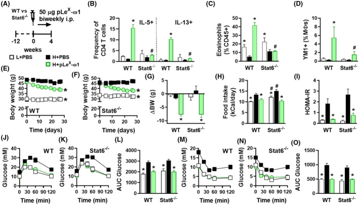
FIGURE 4.

pLeX‐ω1 improves metabolic homeostasis in obese mice by a STAT6‐independent mechanism. WT and Stat6 −/− mice were fed a LFD (white bars) or a HFD for 12 weeks and next received biweekly intraperitoneal injections of PBS (black bars) or 50 µg pLeX‐ω1 (green bars) during 4 weeks (A). At the end of the experiment, eWAT was collected, processed, and analyzed as described in the legend of Figure 1. The frequencies of cytokine‐expressing CD4 T cells (B) were determined. Abundances of eosinophils (C) and YM1+ macrophages (D) were determined. Body weight (E‐G) and food intake (H) were monitored throughout the experimental period. HOMA‐IR at week 4 (I) was calculated, and intraperitoneal glucose (J‐L) and insulin (M‐O) tolerance tests were performed as described in the legend of Figures 1 and 3. Results are expressed as means ± SEM. *P < .05 vs HFD, # P < .05 vs WT (n = 3‐5 mice per group)
