FIGURE 1.

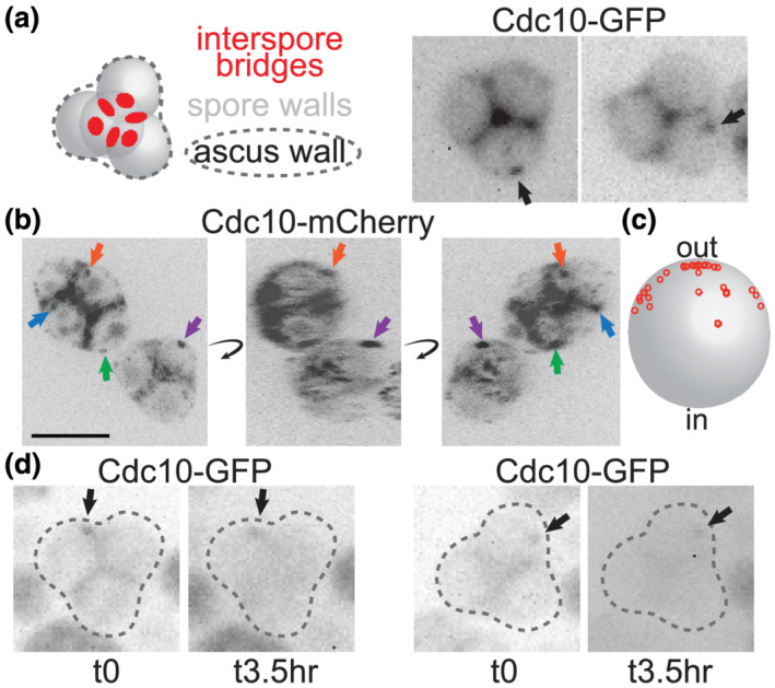
Stable cortical foci of the septin Cdc10 persist on the periphery of asci. (a) At left is an illustration of a typical pyramidal four‐spored ascus in which the three spores at the ‘base’ of the pyramid are in the same focal plane. Red circles indicate interspore bridges. At right, Cdc10‐GFP fluorescence in intact asci from sporulation culture. Arrows indicate cortical foci. Cells are of a diploid strain made by mating haploid strains JTY3985 and FY2742. (b) 2D images from 3D reconstruction of images captured via confocal microscopy of Cdc10‐mCherry fluorescence in asci from sporulation medium. The 3D reconstruction was rotated (curved arrows) to improve visualization of cortical signal versus signal in between spores, which we assume to be in the residual ascal cytoplasm. Arrows point to cortical foci and are color‐coded to track the same foci through the rotation. Scale bar, 5 μm. Cells are of a diploid strain made by mating haploid strains JTY3992 and FY2839. (c) The approximate positions of 35 foci visualized as in (b) were plotted as small red circles onto a large grey circle representing a spore, where ‘out’ refers to the periphery of the ascus and ‘in’ refers to the centre. All foci were on the spore periphery; red circles not at the edge of the grey circle would project into or out of the plane of the image. These data also include foci from diploid cells as in (b) but carrying the CDC10‐mCherry plasmid pZL02. (d) As in (a), but the same asci were visualized before and after a 3.5‐h interval on solid sporulation medium
