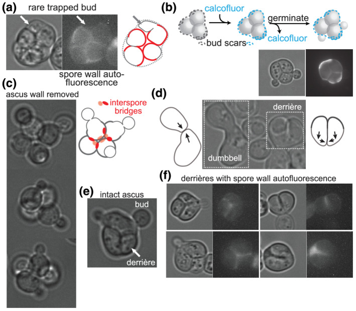
FIGURE 2.

Saccharomyces spores outgrow away from interspore bridges upon germination. (a) Germinating ascus as viewed by transmitted light and with autofluorescence of the spore wall visualized with an RFP filter. (b) According to the illustration, asci were exposed to the chitin‐binding dye calcofluor white and then, after washing away free dye, allowed to germinate. Pre‐existing bud scars on the ascus wall were deposited during diploid budding events prior to sporulation. Calcofluor fluorescence and transmitted light are shown. (c) Tetrads for which the ascus wall was removed by exposure to Zymolyase prior to germination. In the illustration, red circles are interspore bridges, and the spore wall is thicker than the new, vegetative cell wall. (d) Image taken several hours after asci were allowed to germinate, showing dumbbell‐shaped zygote and derrière‐shaped zygote, with illustrations of presumptive directions of outgrowth prior to fusion. (e) Germinating ascus showing two buds penetrating the ascus wall and the other two spores fused into a derrière. (f) As in (a), after 7.75 h of germination, showing localization of the autofluorescent spore wall with regard to the shape of the derrière
