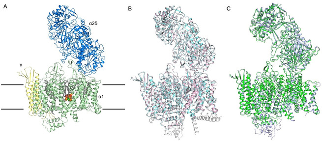
Figure 2.

Nearly identical conformations of rCav1.1 in the presence of DHP compounds in detergents and in nanodiscs. A) Overall structure of the amlodipine‐bound rCav1.1 complex (rCav1.1‐100A) at 2.9 Å resolution. The overall structure of the channel complex is colored for different subunits. The β1 subunit is omitted throughout the manuscript because of its poor resolution. Amlodipine is shown as orange spheres and the bound lipids are shown as gray sticks. The approximate position of the membrane is indicated by black lines. B) The structures of rCav1.1‐100A (with 100 μm amlodipine, pink) and rCav1.1‐100R (with 100 μm RBK, cyan) in nanodiscs and rCav1.1‐200N (with 200 μm nifedipine, gray) in digitonin are nearly identical. C) Structures of rCav1.1‐100S (with 100 μm SBK) in nanodisc (green) and in digitonin (light purple) are nearly identical.
