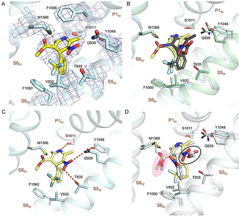
Figure 4.

Coordination of the antagonistic RBK by rCav1.1. A) The densities for the RBK binding site in rCav1.1‐100R. RBK is shown as yellow sticks. The densities, shown as blue mesh, are contoured at 7σ and prepared in PyMol. B) Overlapping binding site for RBK and SBK. The structures of rCav1.1‐100R (blue) and −100S (green) are superimposed. RBK and SBK are colored yellow and black, respectively. C) Coordination of RBK. Potential H bonds are shown as dashed lines. D) Comparison of RBK and nifedipine coordination. The structure rCav1.1‐100R (yellow) and nifedipine (gray) are superimposed. The NO2 group in RBK is highlighted with a black circle. The C3‐ester groups of RBK and nifedipine are highlighted a with pink shadow.
