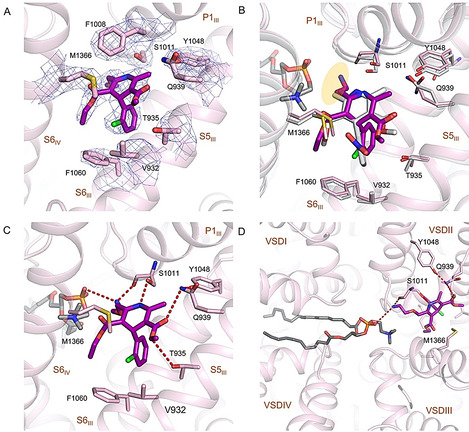
Figure 5.

Specific interactions between rCav1.1 and amlodipine. A) The densities for amlodipine binding site in rCav1.1‐100A. Amlodipine is shown as magenta sticks. The densities, shown as blue mesh, are contoured at 7σ and prepared in PyMol. B) Comparison of amlodipine (magenta) and nifedipine (gray) coordination. The ethanolamine group in amlodipine is highlighted with a yellow shadow. C) Coordination of amlodipine by rCav1.1. The coordinating residues are shown as pink sticks. Potential H bonds are indicated by dashed lines. D) A transverse lipid in the central cavity interacts with amlodipine. The phosphate group of the lipid, assigned as a phosphatidylcholine, and Ser1011 coordinate the ethanolamine group of amlodipine from two opposite sides. Please refer to Figure S5 B in the Supporting Information for the density of the lipid.
