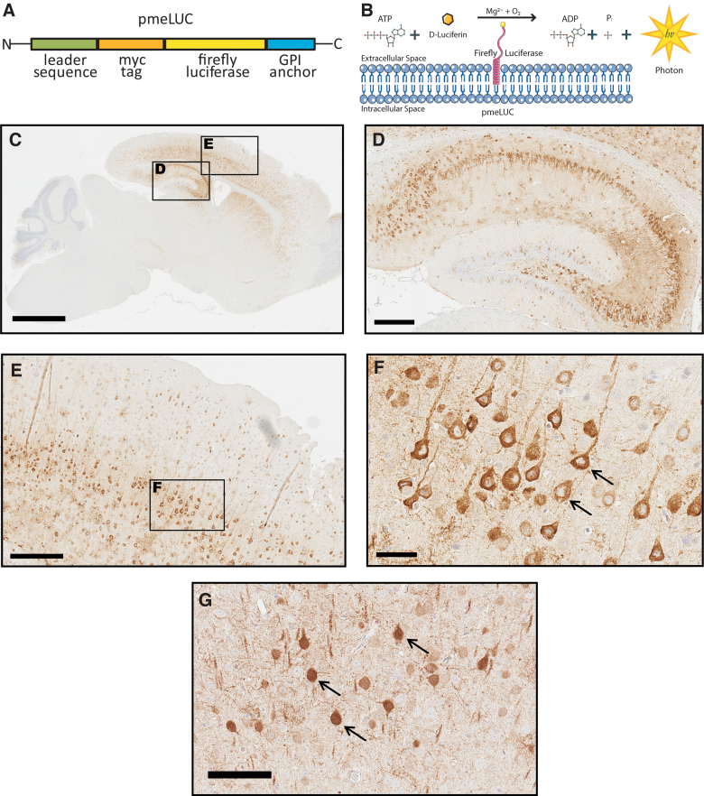
FIG. 1.
(A) Schematic of plasma-membrane–bound pmeLUC construct (B). The enzymatic domain of luciferase is exposed to the extracellular space. Luciferase catalyzes the conversion of D-Luciferin to a photon of light in the presence of ATP. (C) Sagittal mouse brain section immunostained with an anti-firefly luciferase antibody reveals widespread CNS expression of pmeLUC 1 month after injections of pAAV9-CBA-pmeLUC-WPRE into mouse pups. (D, E) Magnified inserts from (C) show pmeLUC expression throughout the hippocampus and cortex. (F) Higher magnification of pmeLUC-positive neurons in the cortex reveals restricted plasma membrane. (G) Control animal injected with pAAV9-CBA-FLuc-WPRE (FLuc) reveals expression throughout the entire neuron, including the nucleus (black arrows). Scale bars, C = 2 mm; D,E = 300 μm; F = 50 μm; G = 100 μm. ADP, adenosine diphosphate; ATP, adenosine triphosphate; CNS, central nervous system; GPI, glycosylphosphatidylinositol.

