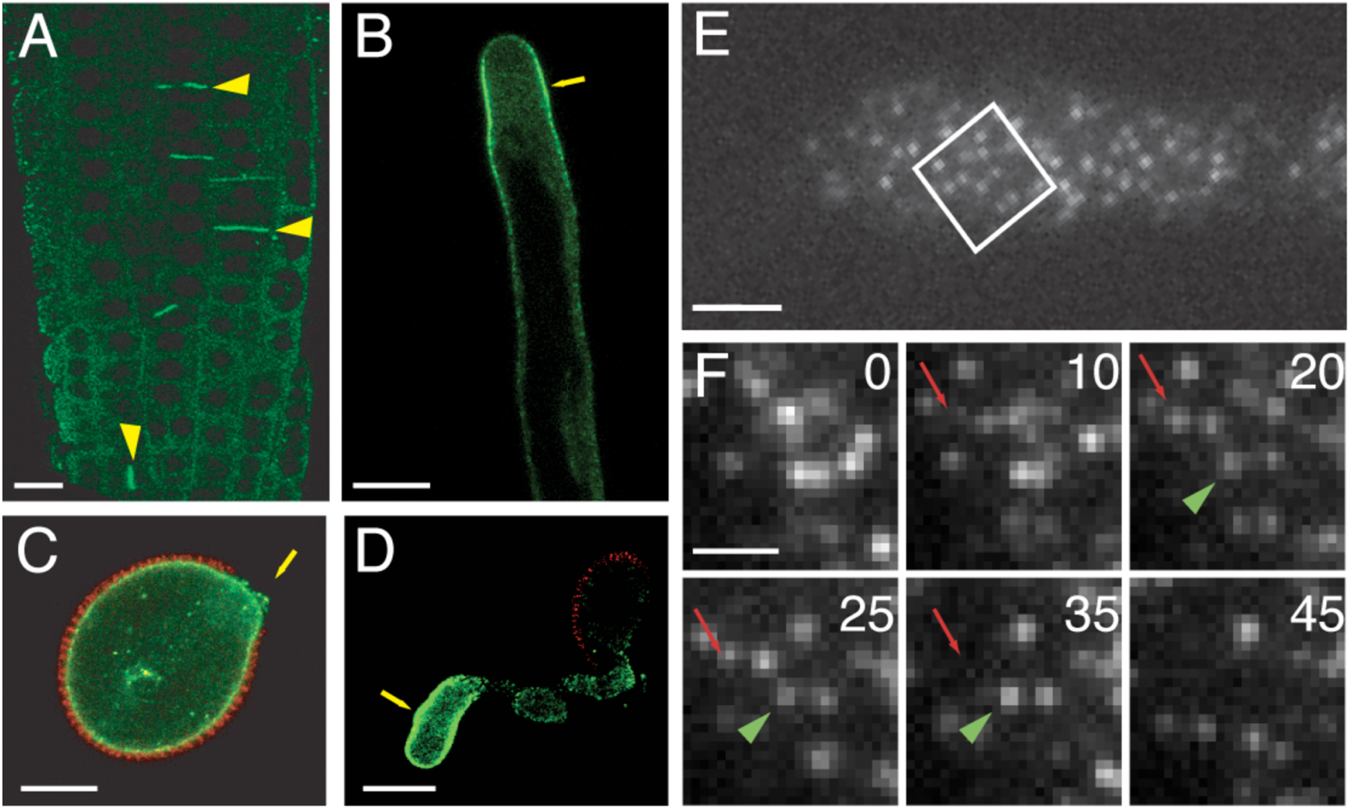
Figure 4. DRP1-FFPs localize to regions of active membrane trafficking.

(A, B) Laser scanning confocal image (LSCM) of DRP1C-GFP (green) in cell plate of dividing cortical root cells (yellow arrowheads) and at the subapical PM (yellow arrow) of an expanding root hair. DRP1A and DRP2A and B show a similar localization at the cell plate in dividing Arabidopsis root cells. (C, D) Pollen from a drp1C-1/DRP1C-GFP plant imaged with LCSM. DRP1C-GFP (green) and autofluorescence of pollen coat (red) are evident. DRP1C-GFP is present at the PM at the aperture of the germinating pollen grain (C) and at the distal end of the pollen tube (D). (E, F) DRP1C-GFP expressing root hair PM imaged with VAEM showing protein in discreet dynamic foci. Time series of boxed area in with two foci indicated with arrow (red) and arrowhead (green) that appear gradually and then vanish over time course (E). Numbers indicate elapsed time from start of imaging in seconds. Bars = 10 μm (A–D), 2 μm (E) and 1 μm (F). Images reprinted from [51] www.plantcell.org “Copyright American Society of Plant Biologists.”
