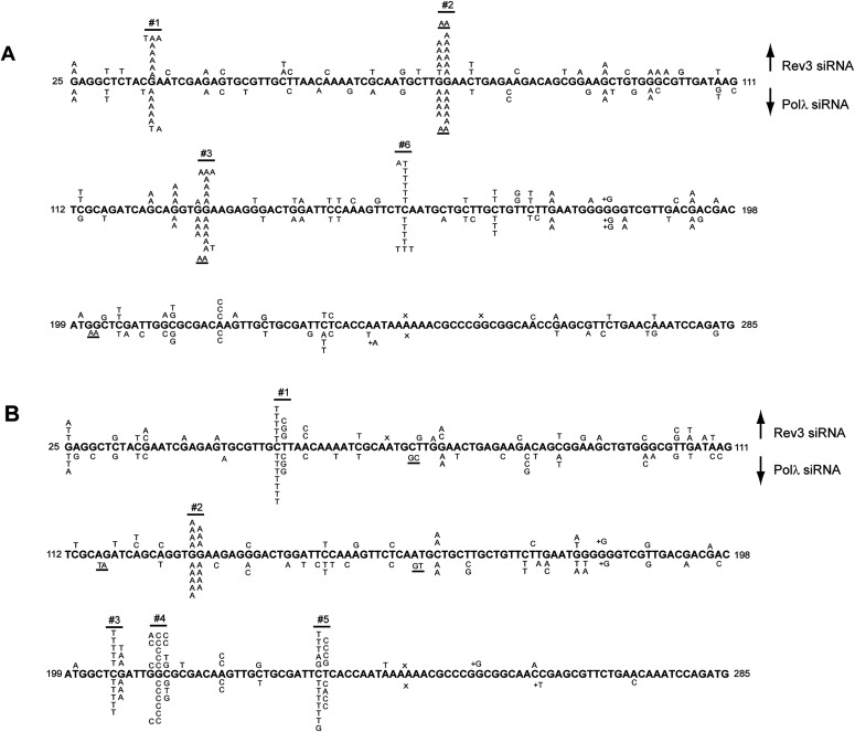
Figure 3. Effects of Polλ depletion on UV-induced mutational spectra in the cII gene in BBMEFs.
(A) UV-induced (5 J/m2) mutational spectra resulting from TLS through cyclobutane pyrimidine dimers (CPDs) in BBMEFs expressing (6-4) PP photolyase. Mutational spectra in Rev3-depleted cells are shown above the sequence and in Polλ depleted cells are shown below the sequence. Whereas TLS through CPDs in WT cells generates hot spots at position #s 1–11 in the cII gene, only hot spots at position #s 1, 2, 3, and 6 remain in Rev3- or Polλ-depleted cells. (B) UV-induced (5 J/m2) mutational spectra resulting from TLS through (6-4) PPs in BBMEF cells expressing CPD photolyase. Mutational spectra in Rev3 depleted cells are shown above the sequence and in Polλ depleted cells are shown below the sequence. The positions of UV-induced hot spots #1–5 are indicated.

