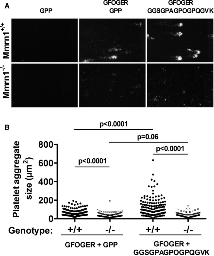
FIGURE 7.

Adhesion of washed, collagen‐related peptide (CRP)‐activated wild‐type and multimerin 1 (Mmrn1)‐deficient mouse platelets to collagen peptides under high shear flow (1500 s−1). A, Representative images show endpoint adhesion of DiOC6(3)‐labeled CRP‐activated Mmrn1+/+ and Mmrn1−/− platelets to immobilized triple‐helical collagen peptides, captured by a Zeiss Axiovert 200 inverted epifluorescence microscope coupled to a AxioCam MRc and Axiovision software (Carl Zeiss Canada Ltd.; original magnification ×20), and (B) mean platelet aggregate size on triple‐helical collagen peptides analyzed for each image captured (n = 5 mice/group, n = 15 images/experiment). Symbols in (B) represent Mmrn1 genotype: +/+ (●) and −/− (x)
