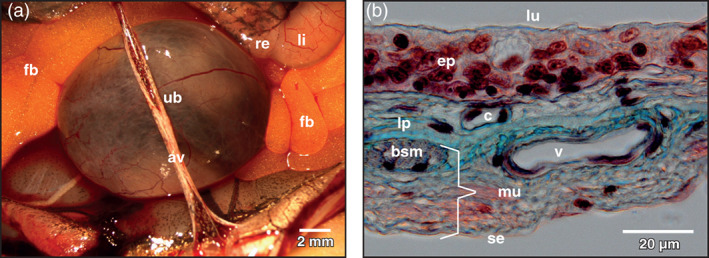
FIGURE 1.

Xenopus laevis, urinary bladder (ub) of an adult individual. (a) Ventral view at the spherical body of the exposed organ. Neck region is hidden. Stereomicroscopy. av abdominal vein, fb fat body, li large intestine, re rectum, ub urinary bladder. (b) Light microscopic structure of the wall of the bladder. Detail view. Paraplast embedded tissue section (7 μm). Goldner's trichrome stain. Note the comparatively thick transitional epithelium (ep). bsm bundle of smooth muscle cells, c capillary, lp lamina propria, lu lumen of bladder, mu muscularis, se serosa, v vein
