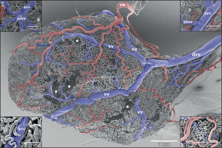
FIGURE 5.

Xenopus laevis, drainage of dorsal and dorso‐lateral areas of the urinary bladder. Dorsal view at the bladder shown in Figure 2(a) . anterior is to the left. Asterisks mark wall areas where incomplete filling of the vasculature occurred. Note four vesical veins (vv) which form the dorsal vesical vein (dvv). Va vesical artery. Inset 1: Formation of a small vesical venule (svv) from postcapillary venules (pcvv). Inset 2: Postcapillary venule (pcvv) draining into a small vesical venule (svv). Note the relation of the calibers of the venules. Inset 3: Capillary (c) draining into a small vesical vein (svv). Inset 4: Undulating capillaries (c) drain in series bilaterally into a postcapillary venule (pcvv)
