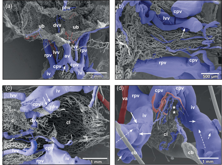
FIGURE 6.

Xenopus laevis, venous drainage of the urinary bladder (ub). (a) Ventro‐caudal view at the large veins running aside the neck region of the bladder. Incomplete specimen. Anterior is at the top, posterior is at the bottom. A lateral vesical vein (lvv) is seen to join the right common pelvic vein (cpv) at its caudal aspect (arrow). (b) Vascular anatomy of the ventral neck region of the bladder after removal of the common pelvic veins (cpv). Anterior is at the left, posterior is to the right. A lateral vesical vein (lvv) is joined by a smaller one (arrow) to finally drain into right or left common pelvic vein (not shown). (c) Vascular anatomy of the cloacal region. Only remnants of the mucosal capillary bed (c) of the dorsal wall of the bladder are left. Note the dorsal vesical vein (dvv) which drains via the posterior hemorrhoidal vein (hv) into the left common pelvic vein (cpv). At its entrance at the caudal margin of the common pelvic vein, the posterior hemorrhoidal vein is broken (arrows). (d) Venous valves of the ischiadic veins (iv). Ventro‐caudal view. In the orthogradely filled large valve, the leaflet structure is clearly replicated (long arrows). Note small valves where retrograde resin flow was stopped at the valves (short arrows). Dashed arrows mark direction of blood flow, asterisks mark slit‐like opening of bladder into cloaca. av abdominal vein, cb conductive bridge, cl cloaca, rpv renal portal vein, va vesical artery
