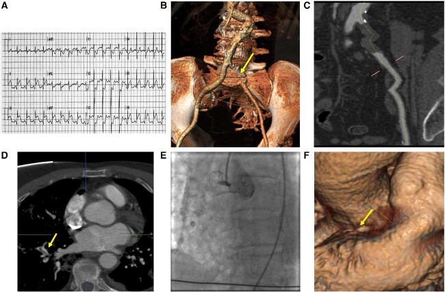
Figure 1.
Electrocardiography, CT and angiographic findings. (A) Electrocardiography showing sinus rhythm with ST-segment elevation in DII, DIII, aVL and V6 and ST-segment depression in V1-V3. (B and C) CT reconstruction of complete left common iliac occlusion. (D and E) evidence at pulmonary CT of thrombus in segmentary arteryE-F: angiographic and CT images of total thrombotic occlusion of right coronary artery.

