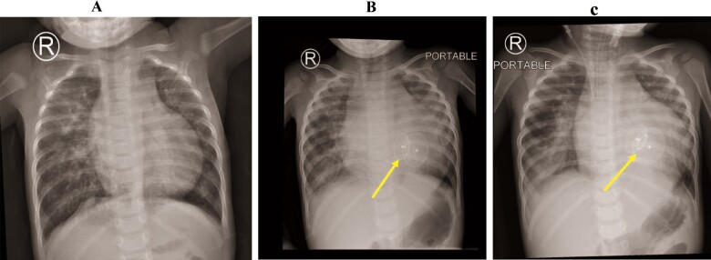
Figure 1.
Chest radiography was conducted before ventricular septal defect closure (A), during the last admission in the emergency department (B), and immediately after mechanical ventilation (C). Results reveal bilateral ground-glass opacities and cardiomegaly. The ventricular septal defect device (yellow arrows).

