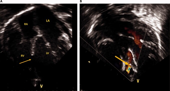
Figure 3.
Echocardiography before and after ventricular septal defect device closure. (A) Apical four-chamber view showing a large mid-muscular ventricular septal defect (yellow arrow). (B) Apical four-chamber view with colour Doppler after device closure showing a small residual leak across the ventricular septal defect device (yellow arrow). LA, left atrium; LV, left ventricle; RA, right atrium; RV, right ventricle.

