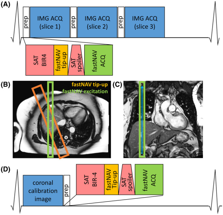
FIGURE 1.

(A) Pulse sequence diagram of cardiac‐triggered CMR perfusion imaging with fastNAV. (B) Geometry of slice‐selective fastNAV pulses overlaid on axial localizer image. The orange rectangle marks the fastNAV tip‐up slice at twice the width of the fastNAV excitation slice (green rectangle). (C) Coronal localizer image showing fastNAV’s frequency encoding direction as blue arrow. (D) Pulse sequence diagram of the fastNAV calibration scan, where fastNAV is acquired after a coronal image of the LV per heartbeat. CMR, cardiac MR; fast NAV, fast navigator; fastNAV ACQ, acquisition of fastNAV using slice‐selective excitation (15°) and frequency‐encoded gradient echo; fastNAV tip‐up, slice‐selective −90° pulse; IMG ACQ, perfusion image acquisition using balanced steady‐state free precession; LV, left ventricle; prep, preparatory pulses for saturation recovery imaging including fastNAV; SAT BIR‐4, saturation with nonselective adiabatic BIR‐490° pulse; SAT spoiler, gradient spoiler to dephase remaining transverse magnetization after saturation pulse
