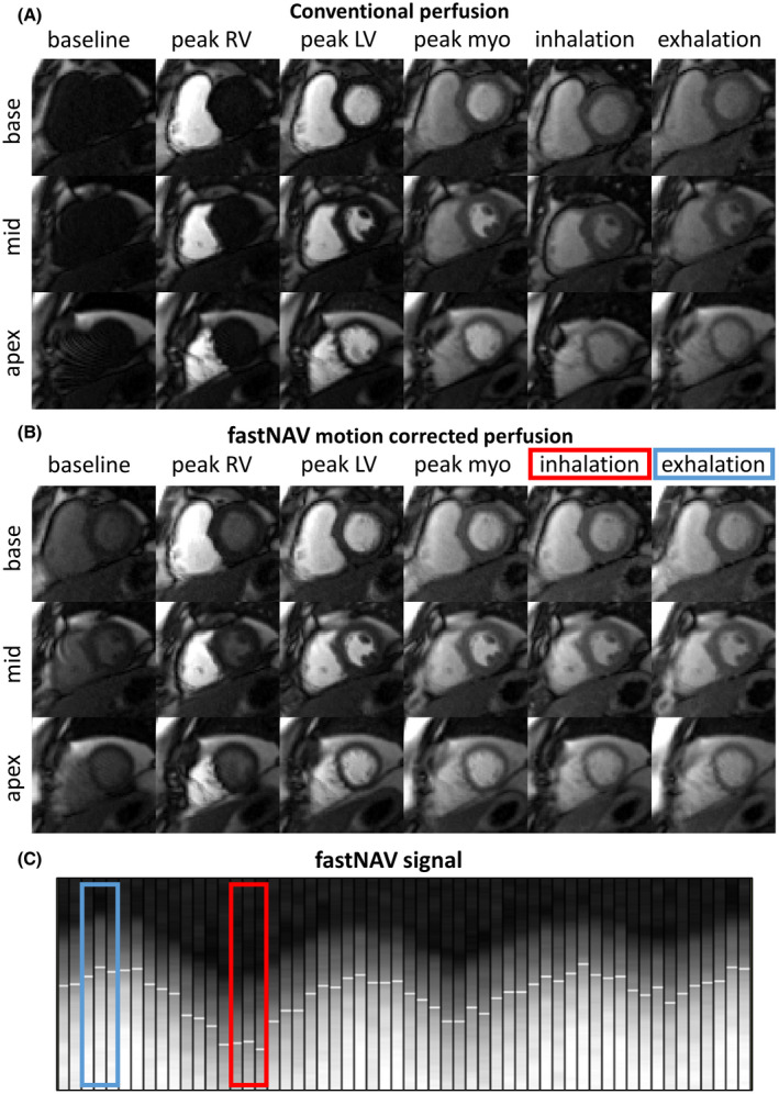
FIGURE 3.

Examples of first‐pass perfusion images acquired in 1 subject (subject 3) under free‐breathing conditions using the conventional and proposed fastNAV approach. Full series are available as video in Supporting Information Videos S1 and S2. Representative dynamics acquired near end‐inspiration and end‐expiration are shown in the last 2 columns. (A) Conventional perfusion without motion correction. Images in end‐inspiration and end‐expiration were selected visually based on the heart’s vertical position. (B) Perfusion sequence with fastNAV motion correction. Images in end‐inspiration and end‐expiration were chosen based on the navigator trace. (C) Segment of the fastNAV signal trace where white horizontal line segments mark the detected navigator positions. The colored rectangles in B and C mark the navigator signal for the images in end‐inspiration (red) and end‐expiration (blue). Substantial motion can be observed between end‐inspiration and end‐expiration images acquired using the conventional sequence. The amount of motion between end‐inspiration and end‐expiration images was substantially reduced using the proposed fastNAV approach. LV, left ventricle; myo, myocardium; RV, right ventricle
