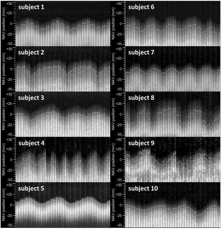
FIGURE 4.

Signal examples of fastNAV acquired during perfusion imaging with fastNAV‐based motion correction for all subjects. Consecutive fastNAV signals acquired over the first 19 heartbeats of each dynamic acquisition (3 fastNAV signals per heartbeat;ie, 1 per slice) are shown for each subject. The fastNAV signal had high SNR and was consistent over time. The visual displacements observed in the fastNAV signal traces were successfully tracked (white horizontal marks)
