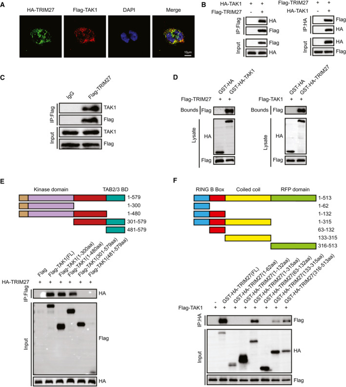
FIG. 7.

TRIM27 interacts with TAK1 through its C‐terminal coiled coil and RFP domain. (A) Representative confocal images show L02 cells transfected with HA‐tagged TRIM27 and Flagged TAK1. Proteins were visualized using anti‐Flag (red) and anti‐HA (green) antibodies, followed by fluorophore‐conjugated secondary antibodies after 24 hours of transfection. Nuclei were stained using DAPI (blue). n = 3 independent experiments per group with six images per group. Scale bar, 25 μm. (B) Flag‐tagged TRIM27 and HA‐tagged TAK1 plasmids were cotransfected into HEK293T cells. Anti‐Flag antibody (left panel) or anti‐HA antibody (right panel) were used for IP. Representative of three independent experiments. (C) Flag‐tagged TRIM27 was transfected into L02 hepatocytes. Anti‐Flag antibody was used for IP. Representative of three independent experiments. (D) Flag‐tagged TRIM27 and GST‐HA–tagged TAK1 plasmids or Flag‐tagged TAK1 and GST‐HA‐TRIM27 were cotransfected into HEK293T cells. Representative of three independent experiments. (E) Full‐length HA‐TRIM27 and various truncated forms of Flag‐TAK1 were cotransfected into HEK293T cells. An anti‐Flag antibody was used for IP. Representative of three independent experiments. (F) Full‐length Flag‐TAK1 and various truncated forms of GST‐HA‐TRIM27 were cotransfected into HEK293T cells. An anti‐HA antibody was used for IP. Representative of three independent experiments. Abbreviations: aa, amino acids; BD, binding domain; DAPI, 4′,6‐diamidino‐2‐phenylindole; IgG, immunoglobulin G.
