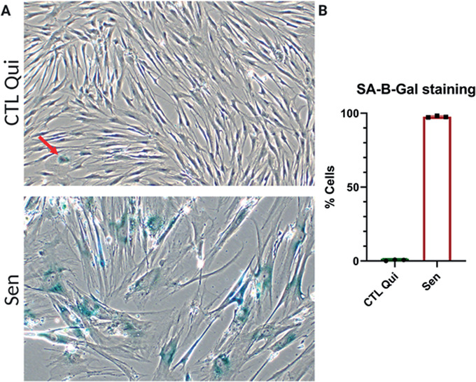
Figure 3.

Senescence‐associated β‐galactosidase (SA‐β‐gal) staining. (A) Representative images of quiescent control (top) and ionizing radiation (IR) senescent IMR‐90 cells (bottom). (B) Quantification of SA‐β‐gal positive cells for quiescent control and senescent cells. Data shown are means of 3 replicates ± SD.
