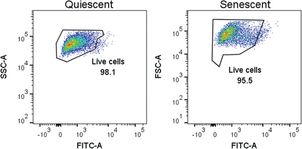
Figure 5.

Propidium iodide (PI) staining for cell death. Percentage of PI‐positive cells in quiescent control (left) and ionizing radiation (IR)‐induced senescent (right) IMR‐90 cell populations. Data shown are representative of both cell populations.
