
FIGURE 6.
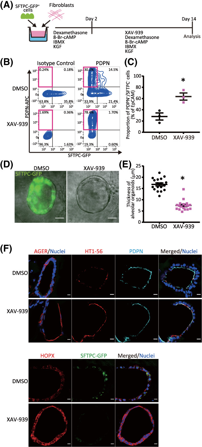
XAV‐939 increases the population of iAT1 cells in FD‐AOs. A, A schematic diagram of the analysis of the effect of XAV‐939 on FD‐AOs. B,C, Flow cytometric analyses of the PDPN and SFTPC expression of FD‐EpCAM+ cells (P2‐5). FD‐AOs (P2‐5) were treated with 10 μM XAV‐939 or DMSO from day 2 to 14, and then the proportion of SFPTC−PDPN+ cells in FD‐AOs was measured on day 14 (mean ± SEM, n = 3 from three independent experiments). The values indicate the percentage of each population. *P < .05 vs DMSO‐treated hiPSC‐derived SFTPC−PDPN+ cells (unpaired two‐tailed Student's t test). D,E, Representative bright‐field and SFTPC‐GFP fluorescence microscopic live cell images of FD‐AOs (P2‐5) treated with DMSO or 10 μM XAV‐939 from day 2 to 14. Thickness of each FD‐AO (P2‐P5) treated with DMSO or 10 μM XAV‐939 was measured (mean ± SEM, n = 18 spheres from three independent experiments). Scale bars = 50 μm. *P < .05 vs DMSO‐treated FD‐AO (unpaired two‐tailed Student's t test). F, Immunostaining of AT1 markers (AGER, HOPX, HT1‐56, and/or PDPN) and SFTPC‐GFP in DMSO‐ or XAV‐939‐treated FD‐AOs (P3). Scale bars = 10 μm. DMSO, dimethyl sulfoxide; FD‐AOs, fibroblast‐dependent alveolar organoids; hiPSC, human induced pluripotent stem cell