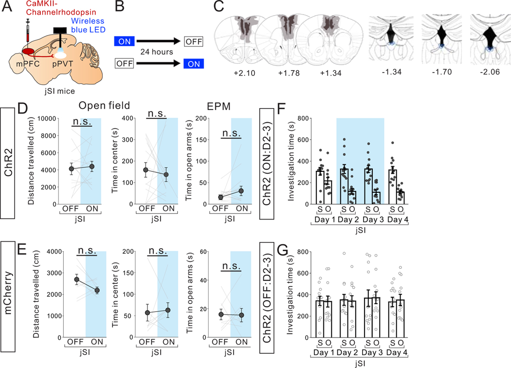
Extended Data Fig. 9. Optogenetic stimulation of mPFC->pPVT projection terminals does not change motor activity or anxiety-related behaviors in adult jSI mice.
(A) CaMKII-ChR2 AAV1 was injected into the mPFC and a wireless blue LED was inserted above the pPVT in jSI mice. (B) jSI mice underwent testing in the open field and elevated plus maze (EPM) with (ON) or without (OFF) light stimulation (20Hz) (C) (left) Viral spread validation from behavior-tested mice. Gray areas represent the minimum (lighter colour) and the maximum (darker colour) spread of ChR2 expression into the mPFC. (right) Optic fiber tip location (blue line circles) was validated in all mice. Experimental images were obtained from 13 mice, three images per mouse for both mPFC and pPVT, with similar results obtained. (D) ChR2+jSI mice showed no differences in motor activity or anxiety-related behaviors between ON and OFF sessions (Left; two tailed paired t-test, t12=0.346, P=0.735, n=13 biologically independent jSI mice, Middle; two tailed paired t-test, t12=0.431, P=0.674, n=13 biologically independent mice, Right; two tailed paired t-test, t12=1.364, P=0.198, n=13 biologically independent jSI mice). (E) Control mCherry+ jSI mice showed no difference in motor activity or anxiety-related behaviors between ON and OFF sessions (Left; two tailed paired t-test, Left; t7=0.970, P=0.365, n=8 biologically independent jSI mice, Middle; t7=0.183, P=0.860, n=8 biologically independent jSI mice Right; t7=0.083, P=0.936, n=8 biologically independent jSI mice). (F-G) Investigation time of each stimulus during first 20 mins of 3 chamber testing from Day 1 to Day 4 of ON group (F) and OFF group (G) during the repeated optogenetic stimulation study in Fig. 8EF. Data in D-G are presented as mean +/− s.e.m.

