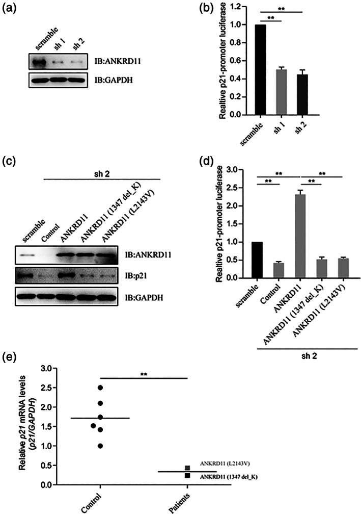
FIGURE 4.

The variants of ANKRD11 attenuate p21. (a) Knockdown efficiency of shRNAs for ANKRD11 by immunoblotting. HEK293T cells were transfected with shRNAs (scramble, sh1 or sh2) for ANKRD11, and the protein levels of ANKRD11 and GAPDH were detected by immunoblotting. Sh1 and sh2 reduced the expression of ANKRD11 protein, respectively. (b) Luciferase reporter assay to detect the relative activities of human p21‐promoter. HEK293T cells were transfected with shRNAs for ANKRD11, as well as with p21‐promoter luciferase reporter and PRL‐TK. The cell lysates subjected to Dual‐Luciferase Reporter assay 48 hrs later. Data are expressed as mean ± SD. **p < 0.01, very significantly difference. With sh1 or sh2, the knockdown of ANKRD11 reduced the p21‐promoter luciferase activities. (c) Detection of the protein levels of p21 by immunoblotting. HEK293T cells were transfected with shRNA (sh 2) for ANKRD11, and reintroduced with wild type ANKRD11, ANKRD11 (p. Lys1347del) or ANKRD11 (p. Leu2143Val). The protein levels of ANKRD11, p21 and GAPDH were detected by immunoblotting. ANKRD11 knockdown reduced the p21 protein expression, while the re‐introduction of wild type ANKRD11 could restore the p21 levels, but not those two ANKRD11 mutants. (d) The variants of ANKRD11 comprised its activating effect on p21 promoter activity. HEK293T cells were transfected with shRNA (sh 2) for ANKRD11, as well as with p21‐promoter luciferase reporter and PRL‐TK, and reintroduced with wild type ANKRD11, ANKRD11 (p. Lys1347del) or ANKRD11 (p. Leu2143Val). The cell lysates subjected to Dual‐Luciferase Reporter assay 48 hrs later. Data are expressed as mean ± SD. **p < 0.01, very significantly difference. Wild type ANKRD11 activated the p21‐promoter luciferase, while those two ANKRD11 mutants lost this function. (e) Comparison of the relative p21 mRNA levels in two patients and healthy controls. The peripheral blood of two patients and six healthy controls were collected. Total RNA was extracted and complementary DNA (cDNA) was synthesized. The relative mRNA levels of p21 were detected by RT‐qPCR. Data are expressed as mean ± SD. **p < 0.01, very significantly difference. Compared with healthy controls, the relative p21 mRNA levels of the two patients were much lower
