
Figure 4.
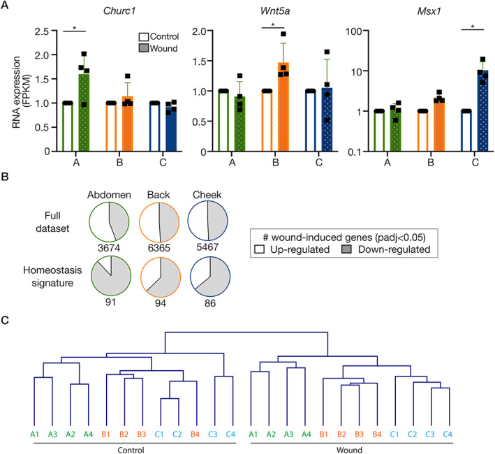
Dermal repair re‐enacts embryonic gene expression programmes. (A) RNA expression (FPKM) of ‘developmental genes’ differentially induced across sites. (B) Pie charts illustrating the directionality of change of all genes significantly altered in wounds versus unwounded samples (padj < 0.05; upregulated: open; downregulated: shaded). Top row: full dataset; bottom row: site signatures in homeostasis from Figure 1. (C) Hierarchical clustering of the samples based on the entire RNA‐seq dataset, showing that the sites remain distinct during a wound response. Abbreviations and colour‐coding: A, abdomen (lateral plate mesoderm‐derived; green); B, back (paraxial mesoderm‐derived; orange); C, cheek (craniofacial, neural crest‐derived; blue).