Figure 3.

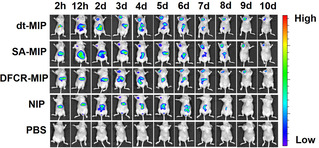
In vivo fluorescence imaging of HepG‐2 tumor (left upper chest) and liver site (upper abdomen) after intravenous injection of NIR797‐doped dt‐MIP, SA‐MIP, DFCR‐MIP, NIP and PBS for different times.

In vivo fluorescence imaging of HepG‐2 tumor (left upper chest) and liver site (upper abdomen) after intravenous injection of NIR797‐doped dt‐MIP, SA‐MIP, DFCR‐MIP, NIP and PBS for different times.