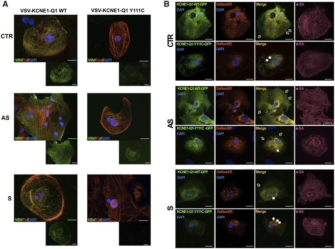
Figure 3.
Trafficking defect of KCNQ1 in iPSC-CMs. (A) Trafficking defect of the VSV-KCNE1-KCNQ1 (VSV-E1-Q1) Y111C fusion protein. Representative images of CTR-, AS-, and S-iPSC-CMs acquired 72 h after transfection with WT or Y111C VSV-E1-Q1 plasmid. Anti-VSV staining (green) was performed on live cells. Then, the cells were fixed, permeabilized, and co-stained with an anti-Troponin-I (TnI) antibody, used as a marker specific for cardiomyocytes (red). Nuclei were stained with DAPI (blue). Scale bar = 20 μm. (B) Retention of Y111C VSV-E1-Q1-GFP fusion protein in endoplasmic reticulum. Images of CTR-, AS-, and S-iPSC-CMs co-transfected with WT or Y111C VSV-E1-Q1-GFP plasmids (green), and pDsRed2-ER plasmid (red). Cardiac cells were visualized by α-sarcomeric actin (α-SA) staining (purple). Nuclei were stained with DAPI (blue). Hollow and filled arrows indicate membrane and ER localization, respectively. Scale bar = 20 μm.

