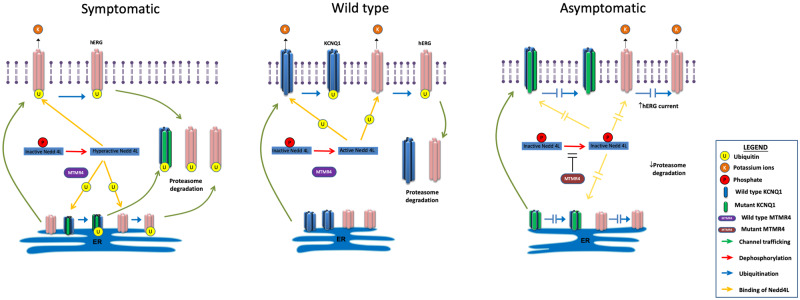
Figure 9.
Schematic illustration of the cellular mechanisms regulated by MTMR4. Cardiomyocytes of normal subject (middle panel): KCNQ1 and hERG are ion channels conducting the IKs and IKr currents, respectively. They are synthetized in the endoplasmic reticulum (ER) from where they traffick to the plasma membrane. Their steady-state expression level at cell surface is physiologically regulated by the ubiquitin ligase Nedd4L. The active form of Nedd4L ubiquitinates ‘old’ KCNQ1 and hERG channels which can then be degraded via the proteasome complex. We have proved that MTMR4 interacts with Nedd4L in iPSC-CMs. Cardiomyocytes of symptomatic patient (left panel): Mutant KCNQ1-Y111C channels are recognized as defective and retained in the ER. This triggers an increased activation of Nedd4L that amplifies the proteasome degradation not only of mutant KCNQ1, but also of WT hERG. Both IKs and IKr are reduced, and repolarization reserve compromised. LQT1 phenotype is manifested. Cardiomyocytes of asymptomatic patient (right panel): SNVs on MTMR4 inhibit Nedd4L activation. The total amount of KCNQ1 and hERG on the plasma membrane is increased compared with cardiomyocytes of the symptomatic patient. IKs deficit is partly compensated by higher IKr expression. LQT1 phenotype is mitigated.

