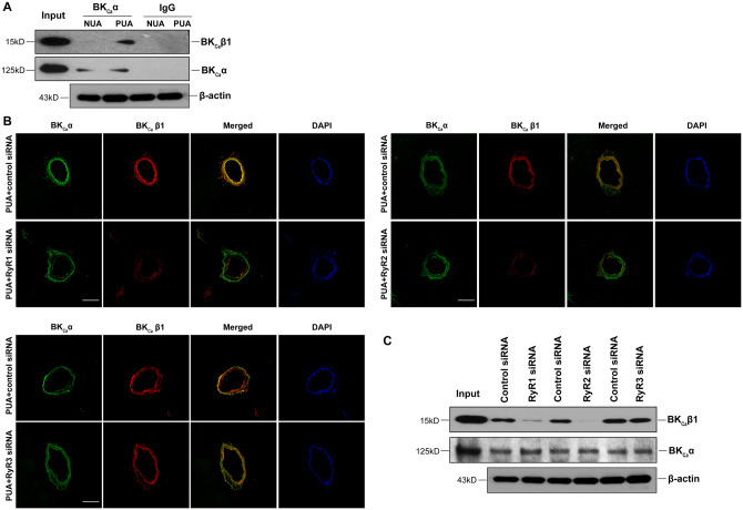
Figure 5.
Knockdown of RyR1/RyR2 reduced the interaction of BKCa channel α and β1 subunits in uterine arteries. (A) Representative immunoblots from five replicates show co-immunoprecipitation of BKCa channel α and β1 subunits in uterine arteries of non-pregnant (NUA) and pregnant (PUA) sheep. Uterine arteries were treated with scramble control siRNA or siRNAs for RyR1, RyR2, and RyR3, respectively, for 48 h. IgG was used as a control to show antibody specificity. (B) Representative confocal immunofluorescence images from five replicates show the co-localization of BKCa channel α and β1 subunits in uterine arteries of pregnant sheep after control siRNAs or RyR siRNAs treatments. The arteries were stained with antibodies against α (green) and β1 (red) subunits. Merged images show in yellow. The nuclear region was stained with DAPI and shows in blue. Scale bar: 100 µm. (C) Representative immunoblots from five replicates show co-immunoprecipitation of BKCa channel α and β1 subunits in uterine arteries of pregnant sheep after control siRNA or RyR siRNAs treatments. β-Actin blots showing equal total protein lysates (input).

