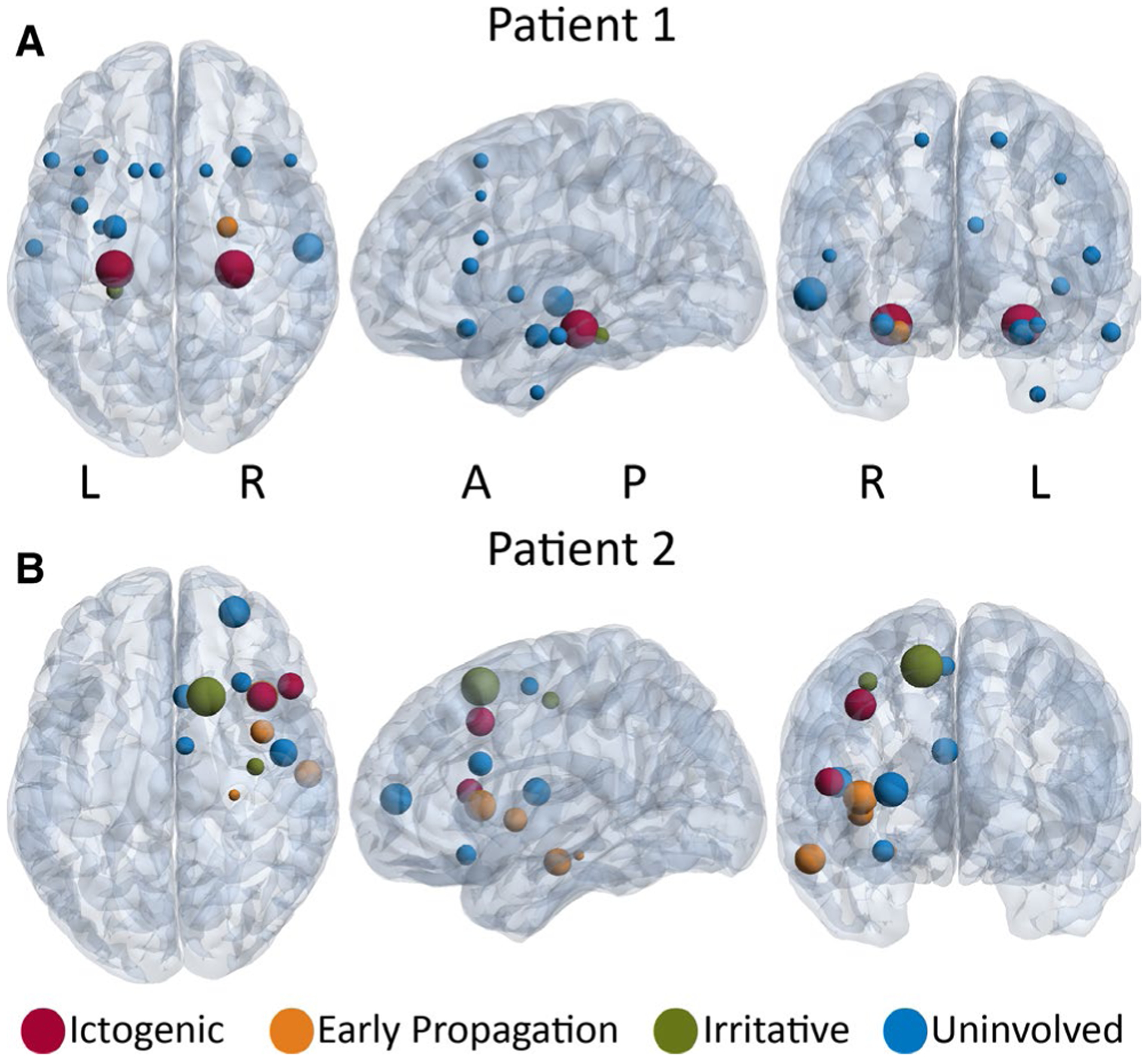
FIGURE 2.

Mutual information (MI) strength maps in two example patients. A, Patient 1 is a right-handed, 29-year-old woman with 8 y duration of epilepsy. The ictogenic regions (red) are the left and right hippocampi. The early propagation region (orange)is the right amygdala, and the irritative region (green) is the left parahippocampal gyrus. In this patient, ictogenic regions demonstrate highest MI strength. B, Patient 2 is a right-handed, 24-year-oldman with 10 y duration of epilepsy. The ictogenic regions (red) are the right inferior frontal gyrus and middle frontal gyrus. The early propagation regions (orange) are the right insula, frontal operculum cortex, hippocampus, and middle temporal gyrus. The irritative regions (green) are the right precentral gyrus and superior frontal gyrus. In this patient, no clear relationship was seen between epileptogenicity and MI strength. In both panels, the size of the spheres is proportional to MI strength of the region. A full list of the regions sampled are listed in Figure 4 legend. A = anterior, L = left, P = posterior, R = right
