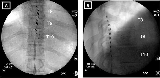
Figure 1.
Radiographical images of implanted percutaneous leads. (A) Anterior/posterior view showing the left lead positioned at the top of T8 with the right lead positioned at the middle of T8. The staggered configuration provides overlap of the T8–T9 disc space and T9–10-disc space corresponding to the targeted dermatomes for low back and leg pain. (B) Lateral view indicating the leads are correctly positioned in the posterior epidural space.

