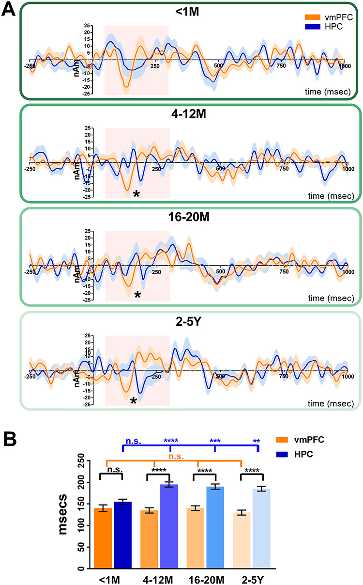
Figure 6.

Initiation of retrieval—the effect of AM remoteness. (A) Event-related signals for AM retrieval and baseline counting for the vmPFC (in orange) and the left hippocampus (in blue). The continuous lines represent the mean, and the shaded areas around the lines represent the SEM. The pink shaded boxes highlight the period from 50 to 300 ms in which the maximum response was examined. * = significant difference between the vmPFC and left hippocampus engagement (with Bonferroni correction at P < 0.01). (B) Bar graph displaying the means and SEM of the maximum responses for AM for the vmPFC (orange bars) and the left hippocampus (blue bars). For AMs <1-month-old, the maximum response of the vmPFC and left hippocampus occurred at around the same time. For all other AM ages, the maximum response of the vmPFC occurred significantly earlier than the left hippocampus. vmPFC = ventromedial prefrontal cortex, HPC = hippocampus, M = months, Y = years, ns = no statistically significant difference, ** = P < 0.01, *** = P < 0.001, **** = P < 0.0001.
