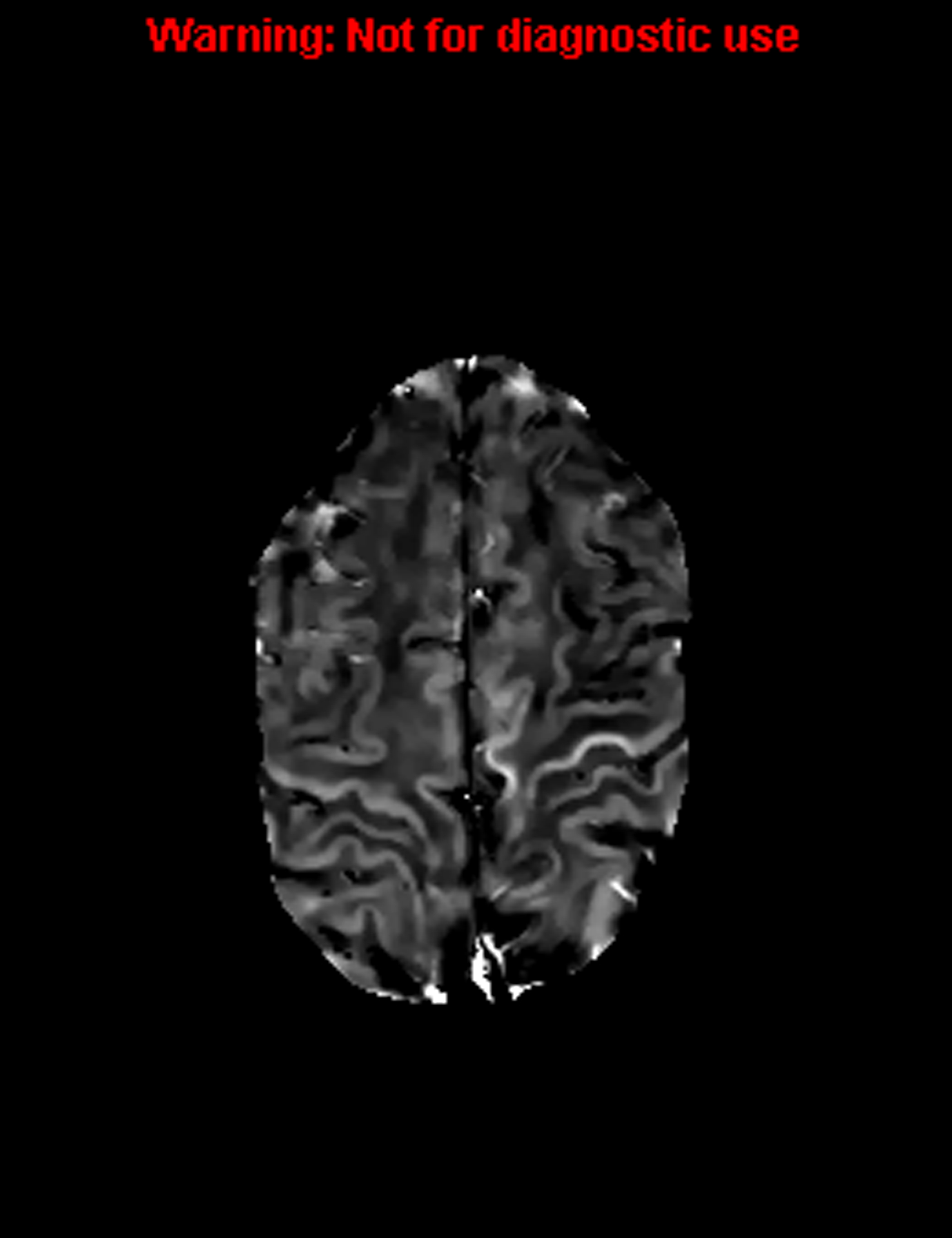
Figure 1. Increased signal in the left arm and leg.

T2 hypo-intensity in PLS patients and T2*/R2* susceptibility measures correlate with microglial iron deposition in the middle and deep cortical layers on autopsy. The changes can be appreciated in specific regions of the homunculus that can be correlated with the clinical syndrome.
