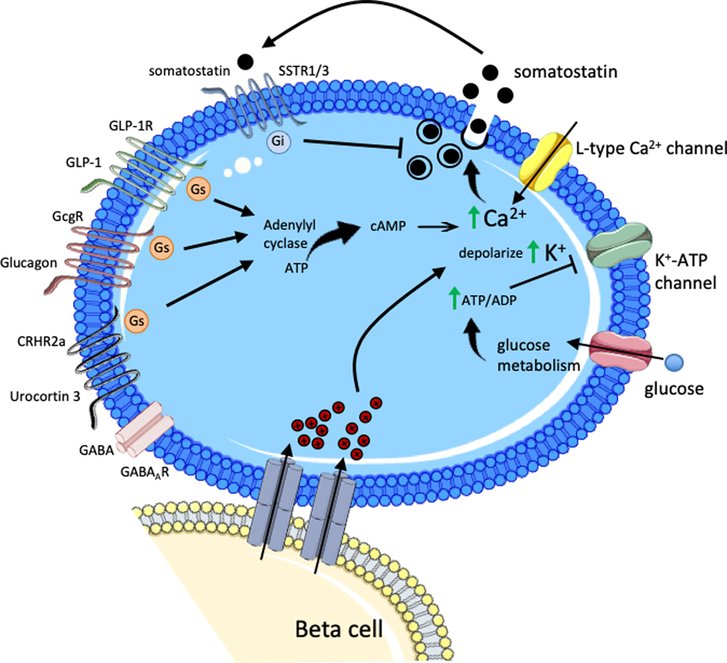
Figure 3. Islet paracrine regulation of delta cell somatostatin secretion.
During hyperglycemia, increasing glucose is taken up by the delta cell and metabolized to generate ATP, which increases the ATP/ADP ratio causing closure of K+-ATP channel. The resulting depolarization by intracellular K+ accumulation stimulates the L-type calcium channel leading to action potential firing and a rise in intracellular calcium that stimulates somatostatin exocytosis. Gap junctions connecting beta cells and delta cells allows transmission of electrical signals from beta cells induce depolarization and somatostatin secretion in adjoining delta cells. Beta cell secretory molecules Urocortin 3 and GABA, packaged with insulin granules, mediate beta cell action on delta cells. Urocortin 3 acts through the corticotropin-releasing hormone receptor (CRHR) 2a activating the stimulatory Gs protein, which induces adenylyl cyclase. Subsequent elevation of intracellular cAMP and rise in intracellular calcium levels leads to somatostatin exocytosis. GLP-1 and glucagon similarly act through Gs activation of adenylyl cyclase to stimulate somatostatin secretion. Somatostatin auto-/paracrine signaling in delta cells occurs through somatostatin receptor (SSTR) 1/3 activation of inhibitory Gi protein.

