FIGURE 2.

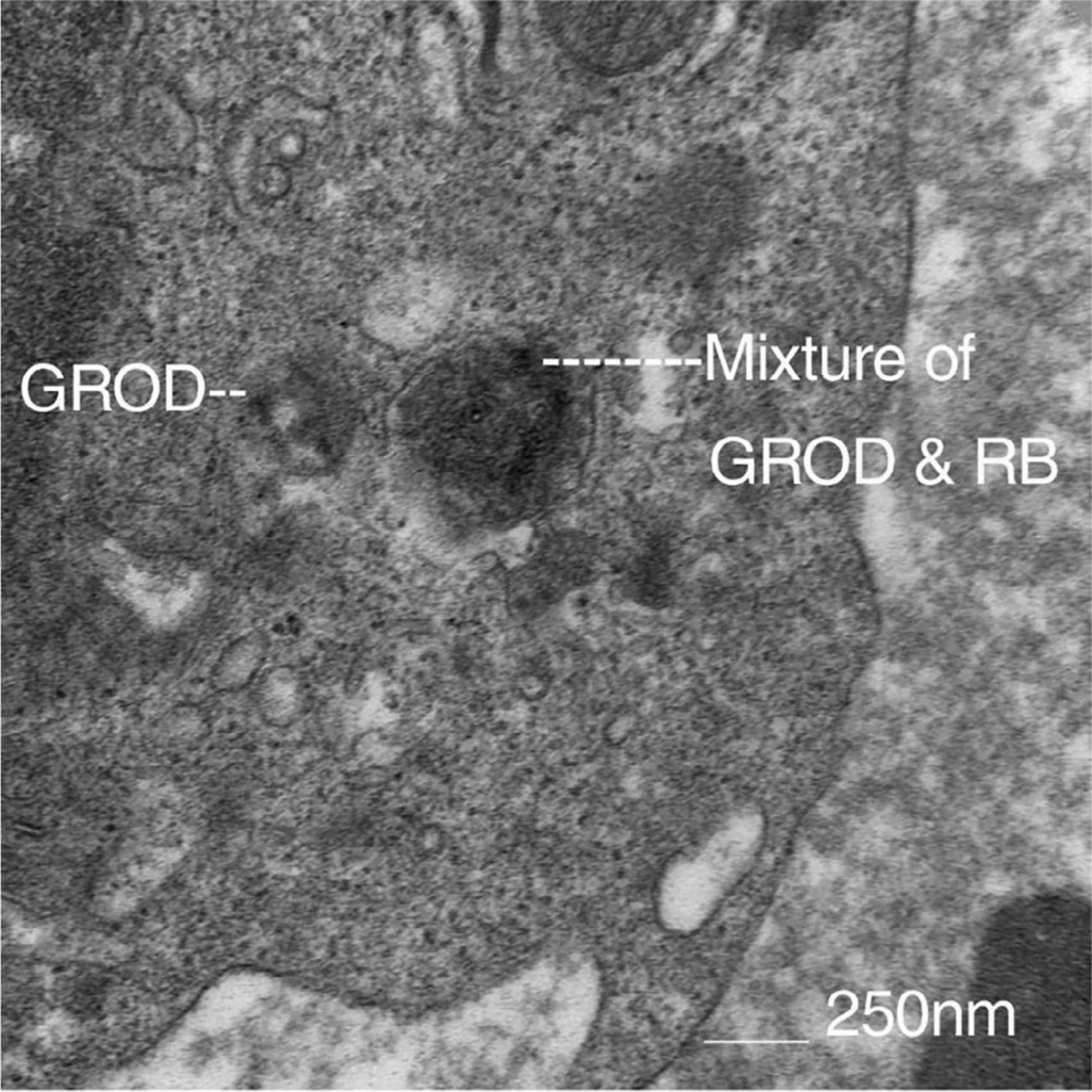
Electron Microscopy study of peripheral lymphocytes of one of our patient’s storage inclusions with morphology of granular osmophilic deposits (GROD) on the left, and GROD and rectilinear body (RB) mixed type on the right

Electron Microscopy study of peripheral lymphocytes of one of our patient’s storage inclusions with morphology of granular osmophilic deposits (GROD) on the left, and GROD and rectilinear body (RB) mixed type on the right